
Looking Inside: The Jackson School’s World Famous CT Lab Gives Us a Peek into Some of the World’s Most Precious Artifacts
October 14, 2016
Rocks from Space
By Monica Kortsha
In June 2016 the Jackson School hosted visitors from other worlds.
Since coming to Earth with astronauts on the Apollo missions or being plucked by scientists from the Antarctic ice, the visitors—moon rocks and meteorites from Mars, the moon and asteroids—have become some of the most studied samples in NASA’s collection.
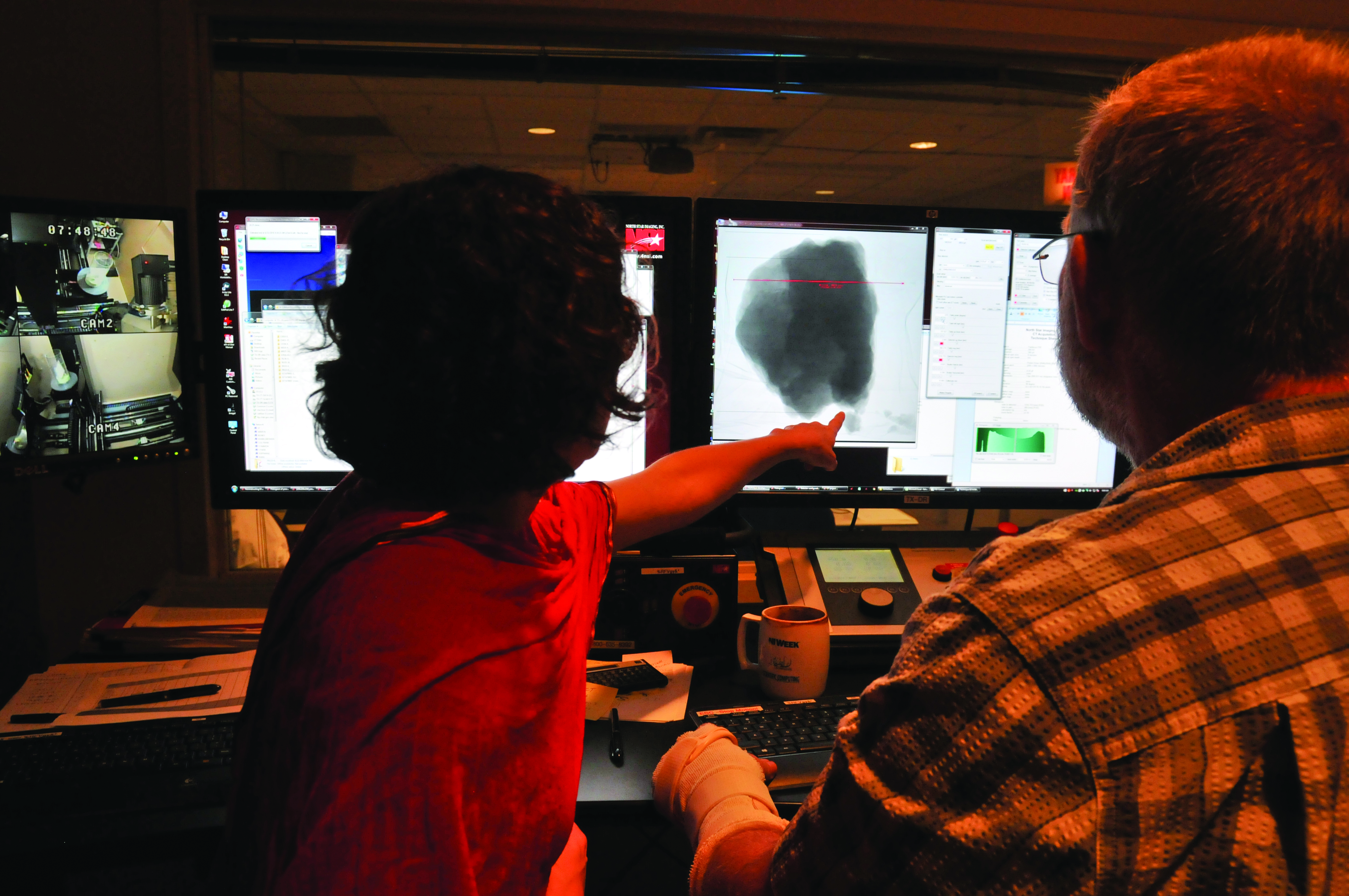
This summer a select few were brought to the Jackson School’s High Resolution X-ray Computed Tomography Facility, or UTCT, to capture a perspective that no one has seen before: a view from the inside.
NASA’s plan is to combine the interior view from CT with high-resolution precision photography to create a 3-D virtual model useful to scientists and space aficionados alike, said Erika Blumenfeld, a visual artist and conservation scientist who is designing the model for NASA.
“Combining these two datasets into a 3-D virtual model— where you can zoom in to see details at 60 microns or greater, rotate it around and really get a sense of the rock—will provide an incredible amount of information,” Blumenfeld said.
“Additionally, we can make lower resolution versions for NASA apps and include the evolving story of the rock’s formation, so kids and adults alike can begin to explore what scientists have learned from these special rocks from space.”
The lab’s scientific CT scanners work like a high-powered clinical CAT scan. Because the samples imaged by the lab are not living, they can be safely scanned with high-energy X-rays that clearly bring features as small as 10 microns (less than the width of a human hair) into view, depending on the size and density of the sample.
The Johnson Space Center in Houston is in the process of buying its own CT scanner to image its thousands of samples. For the first batch of rocks, a collection of 27 specimens, they’re learning the ropes from the experts at the Jackson School.
“These guys have been running the facility for 20 years, so we’re collaborating with the best in the nation here,” said Cindy Evans, Johnson Space Center astromaterials curation chief. The Jackson School’s lab will scan more batches from Johnson Space Center in the future.

UTCT Lab Director Richard Ketcham, a professor and Jackson School Associate Dean for Academic Affairs, has made a name for the lab in scanning and analyzing a wide range of geological specimens, from fossils to crystalline rocks, and also a few meteorites over the years. Romy Hanna, a Ph.D. student at the Jackson School advised by Ketcham, has been helping to further the use of scanning on space rocks. Her own research on carbonaceous chondrites—primitive meteorites made of leftover material from the formation of the solar system—has helped increase UTCT’s repertoire and deepened its involvement with meteorites.
By assisting NASA in scanning its samples, Hanna hopes to showcase the value of CT and further strengthen the bonds between the lab and the planetary geology community.
“CT is interesting in the meteorite world because it hasn’t been used very much,” Hanna said. “We have this amazing technique that can document the interior of these really rare objects … so it’s very desirable that we be more involved in the meteorite community and scan more samples.”
The CT scanning project is a new direction for NASA. But collaborations with the Jackson School go back to the space program’s Apollo days, with Department of Geological Sciences Professor Bill Muehlberger teaching field geology to astronauts and helping define the scientific objectives for the Apollo 16 and 17 missions. Some of the rocks the lab will be scanning include samples brought back from those very missions. However, “Big Muley”—a nearly 26-pound find from Apollo 16 named in Muehlberger’s honor—wasn’t among the samples. As the largest rock ever recovered from the moon, it’s too big to scan in the lab’s instrument.
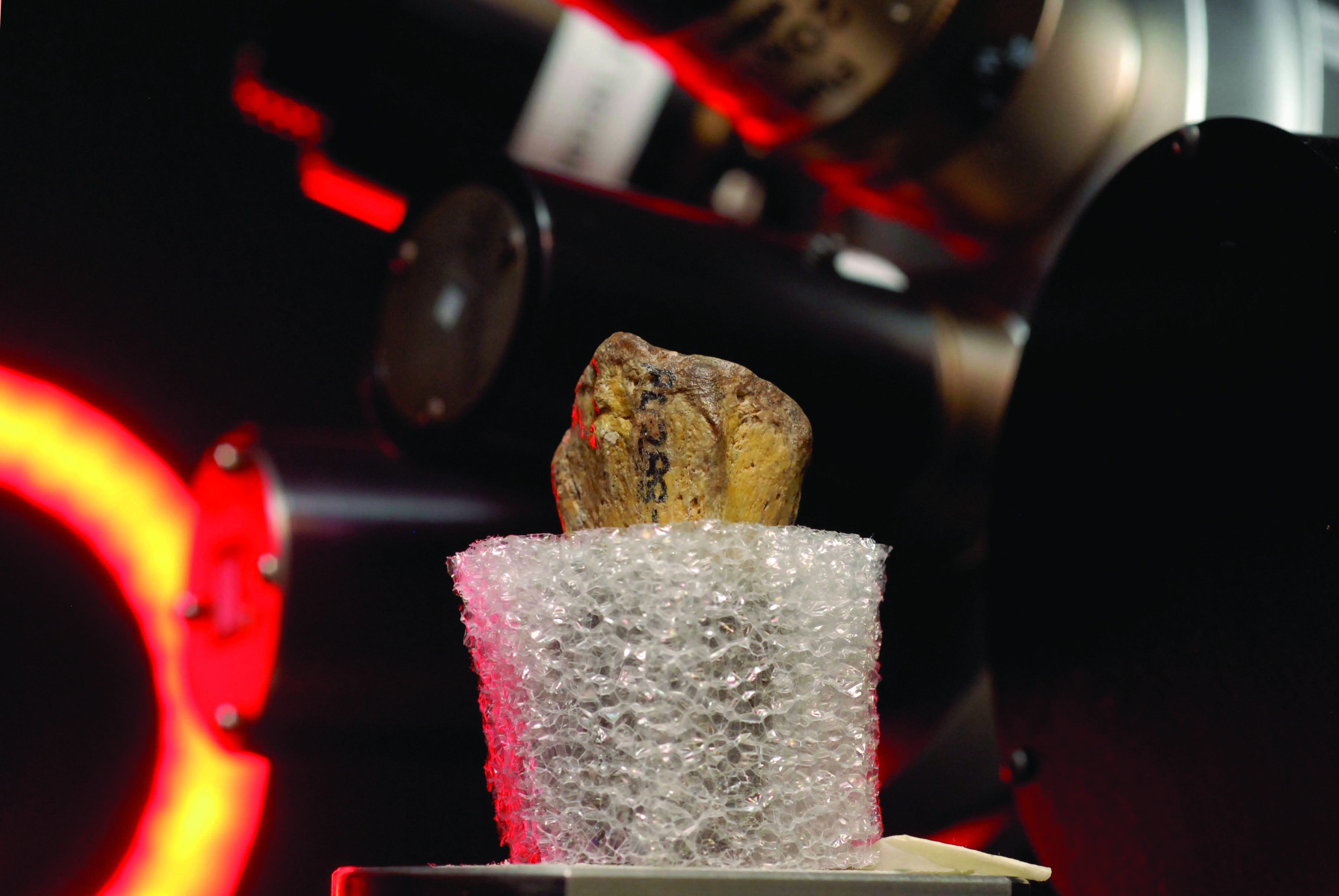
At their home at the Johnson Space Center, the space rocks are secured in nitrogen-gas-filled cabinets within a vault. During their two weeks at UTCT the rocks were kept in the more modest accommodation of a blue Patagonia duffle bag locked in a safe, with each sample sealed inside a silvery opaque “travel case” made up of three nitrogen-filled bags nested inside each other.
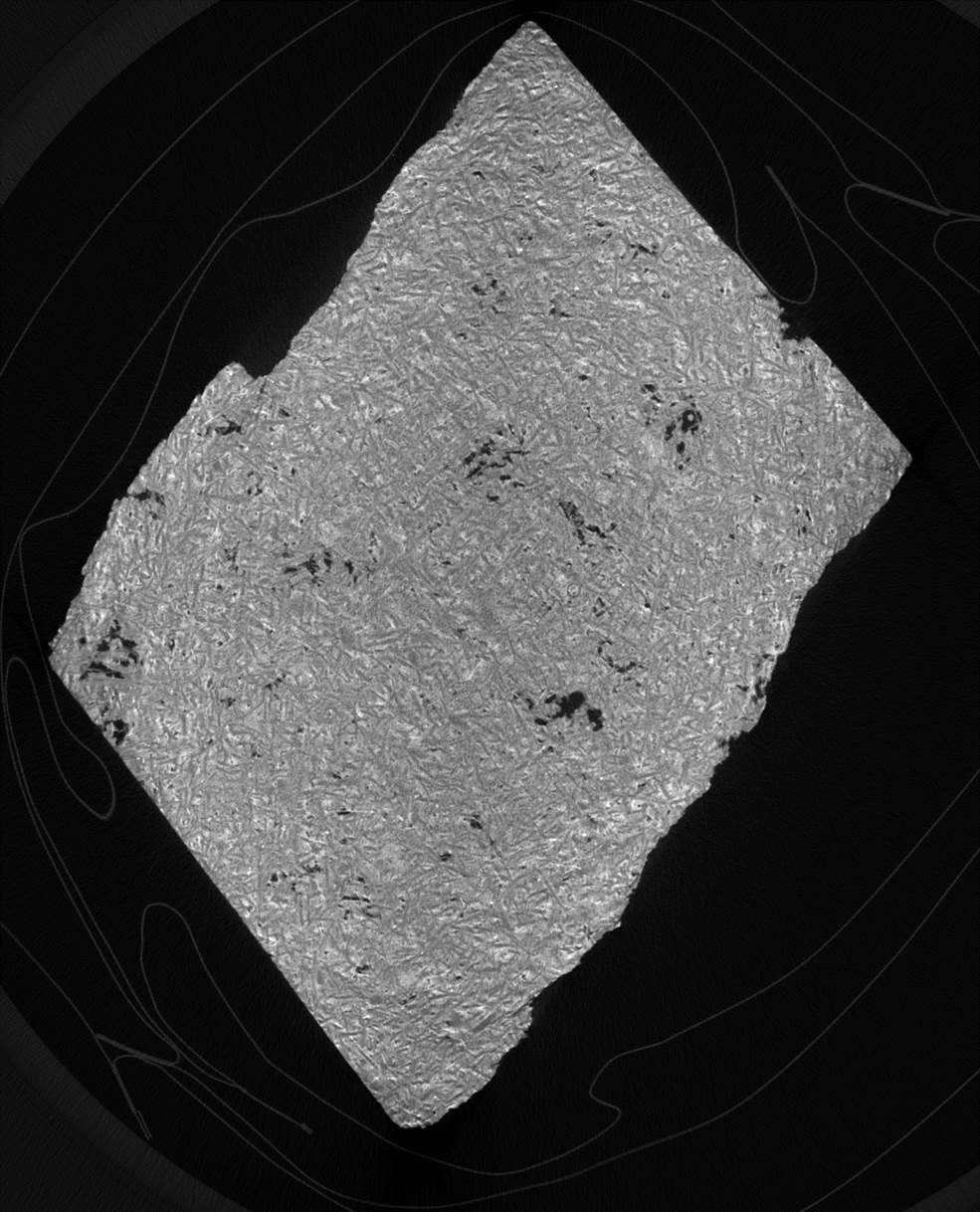
The bags blur the rocks’ features from the outside, but disappear completely in CT images that reveal the individual mineral grains that comprise the celestial material.
Some of the specimens are originally from Vesta, the second-largest and brightest object in the asteroid belt. Others are carbonaceous chondrites, Hanna’s expertise.
From the moon, there are breccias—rocks broken and fused together by the force of an impact like an asteroid—basalts from lunar lava flows, as well as crumbly rocks from the old and cratered highlands.
“Because these samples were chunks on the surface of the moon, they’ve been smashed by multiple impacts and become really friable, crumbly,” Evans said. “So one of the things that we’ll be able to do because of the CT scans is have critical data about the samples without having to handle them as much.”
All the rocks are scientifically important. But a handful of samples were selected primarily for their celebrity. Parts of Lunar basalt 70017, nicknamed the “Children of the World Rock,” served as a goodwill gesture toward humanity when, in 1973, President Richard Nixon had small chunks of the moonrock mounted on plaques and distributed to all countries in the world and to all 50 United States.
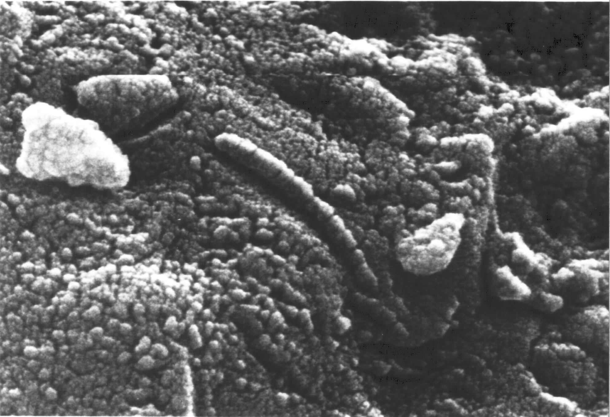
And in 1996 the Martian meteorite Allan Hills 84001 made headlines—and sparked controversy—when NASA announced the rock could hold evidence for life on Mars in the form of microscopic features that looked like fossilized bacteria. The actual identity of the structures remains an open question.
By the end of its collaboration with the Jackson School, NASA plans to scan at least 60 rocks. The CT scans of the first 27 samples will soon provide wider access to rare materials that only a handful of scientists and astronauts have been able to examine up close—and a first look at what’s inside.
Evans said she’s excited to open up the opportunity to examine rocks from the moon, Mars and other worlds to more people.
“It’s challenging for individual students or investigators to get permission to come into the lab, and suit up and interact with the sample, and it’s more challenging to be able to provide that opportunity to a larger number of people for understandable reasons,” she said.
“With the 3-D models, we’ll be able to share samples with people anywhere.”
Solving a 3.2-million-year-old mystery
By Anton Caputo
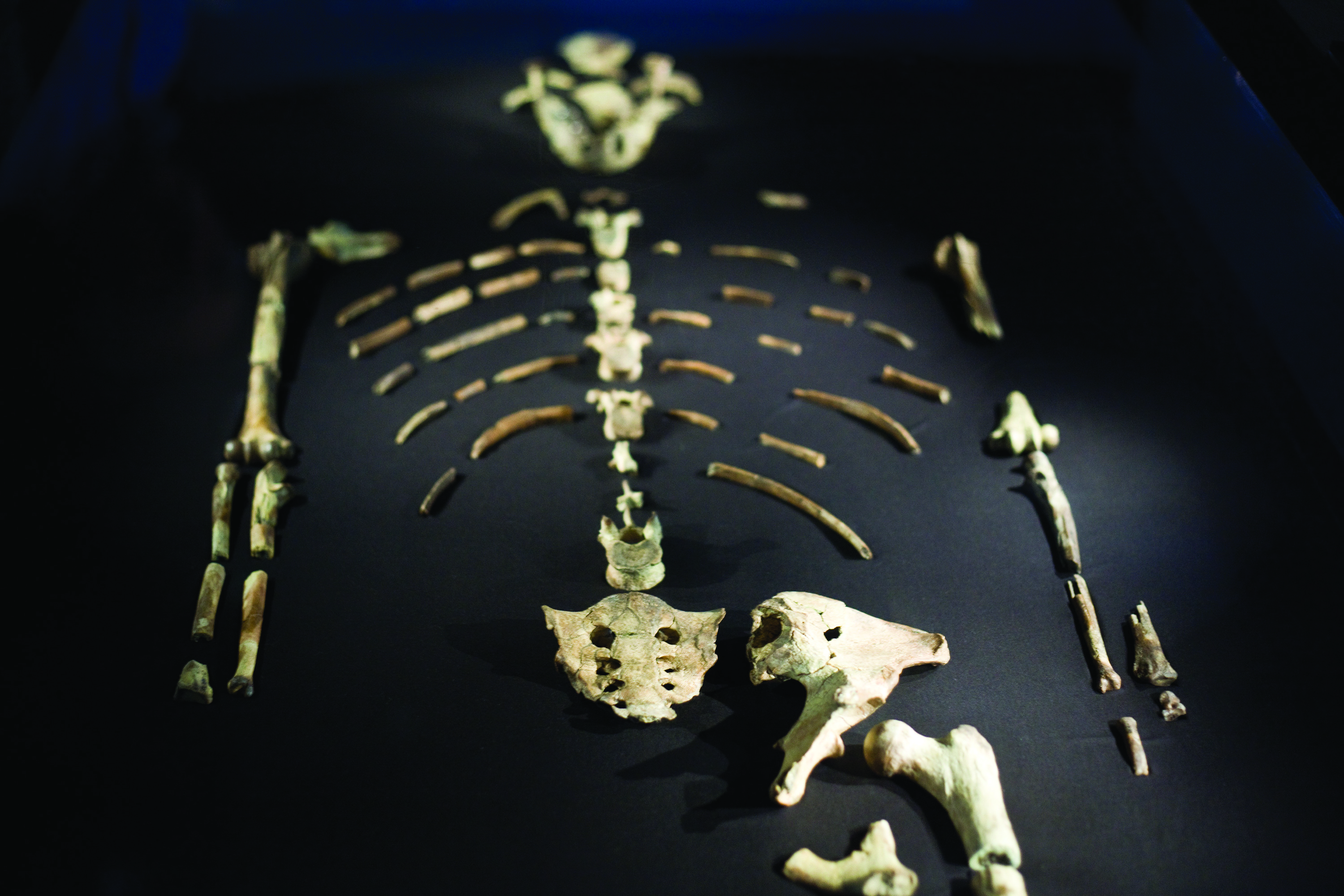
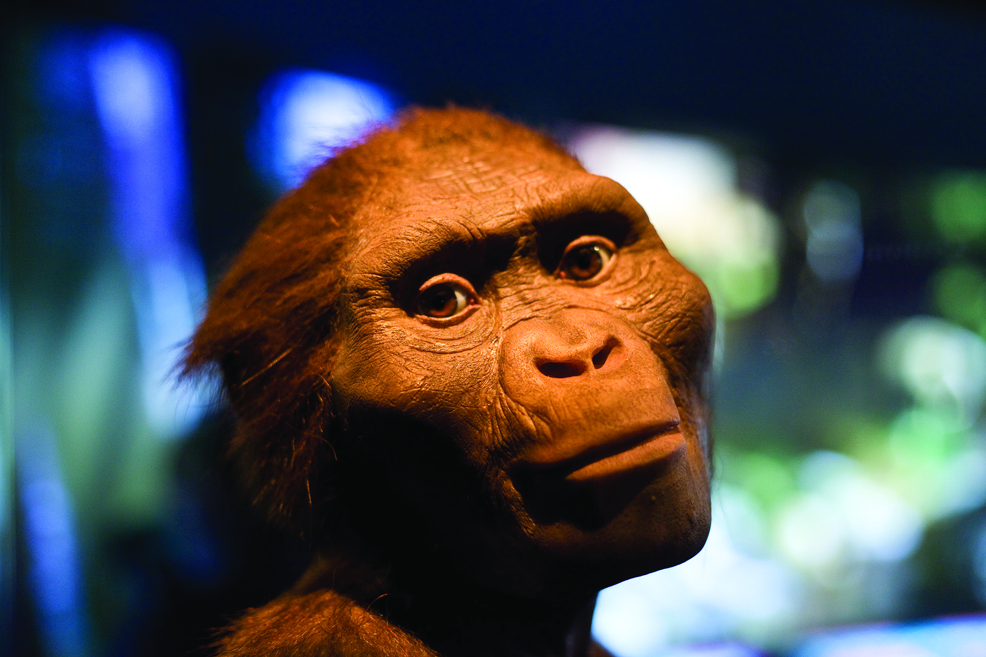
University of Texas researchers closed one of the coldest of cold cases in August 2016 when they revealed that Lucy, the world’s most famous fossil of a human ancestor, likely died after falling from a tree.
The research, published in Nature, was made possible because eight years ago the celebrated fossil took a detour from a U.S. museum tour to the Jackson School’s High-Resolution X-ray Computed Tomography Facility (UTCT).

Over 10 days at the lab in 2008, UT Austin anthropology and geological sciences Professor John Kappelman and geological sciences Professor Richard Ketcham carefully scanned every piece of Lucy’s 40-percent-complete skeleton to create a digital archive of more than 35,000 CT slices. Ketcham, who is also the lab’s director and Jackson School Associate Dean for Academic Affairs, said researchers jumped at the chance to study Lucy, a 3.2-million-year-old specimen of Australopithecus afarensis that is among the oldest, most complete skeletons of any adult, erect-walking human ancestor. The crew prepared to work day and night and bought a safe to keep the priceless fossil secure —a safe they wouldn’t use again until June 2016, when it housed meteorites and Apollo moon rocks from NASA.
The lab’s equipment is designed to scan through solid rock at a higher resolution than medical CT. This made the lab one of the few places in the world capable of scanning something as precious as Lucy.
“We were the first industrial CT scanner in a science department in the world when we got our scanner in 1997, and we were recognized as the premier place to have this kind of work done,” Ketcham said. “There’s only one Lucy, and you want to study her as much as possible. CT is nondestructive, so you can see what is inside, the internal details and arrangement of the internal bones.”
The discovery of the cause of Lucy’s death would come years after the scanning when, studying Lucy and her scans, Kappelman noticed something unusual: The end of the right humerus was fractured in a manner not normally seen in fossils, with the round head of the joint collapsed and driven into the shaft, while preserving a series of sharp, clean breaks with tiny bone fragments and slivers still in place.
“This compressive fracture results when the hand hits the ground during a fall, impacting the elements of the shoulder against one another to create a unique signature on the humerus,” said Kappelman, who consulted Dr. Stephen Pearce, an orthopedic surgeon at Austin Bone and Joint Clinic. Kappelman showed him a modern human-scale, 3-D printed model of Lucy and asked how he would diagnose the break.
Pearce confirmed: The injury was consistent with a four-part proximal humerus fracture, caused by a fall from considerable height when the conscious victim stretched out an arm in an attempt to break the fall. Kappelman observed similar but less severe fractures at the left shoulder and other compressive fractures throughout Lucy’s skeleton including a pilon fracture of the right ankle, a fractured left knee and pelvis, and even more subtle evidence such as a fractured first rib—“a hallmark of severe trauma.”
These marks were all consistent with fractures caused by a fall. Without any evidence of healing, Kappelman concluded the breaks occurred perimortem, or near the time of death.
A question remained: How could Lucy have achieved the height
necessary to produce such a high velocity fall and forceful impact? Kappelman argued that because of her small size—about 3 feet 6 inches and 60 pounds—Lucy probably foraged and sought nightly refuge in trees.
When the findings were announced, news of the research flooded major science and news outlets worldwide. But the theory of her death is not without controversy. That’s because Lucy has been at the center of a vigorous debate about whether this ancient species spent time in the trees in addition to walking on the ground since her discovery in the Afar region of Ethiopia in 1974 by Arizona State University anthropologist Donald Johanson and graduate student Tom Gray.
Thanks to the UTCT and the Ethiopian National Museum, anyone can evaluate the hypothesis for themselves. The researchers have provided access to 3-D files of Lucy’s shoulder and knee for the public to download and print on a 3-D printer.
“We are hoping that releasing these files helps lead to a transformation in the culture of how data like this are handled,” Ketcham said. “For over 30 years, Lucy was kept in a vault in Ethiopia and few were allowed to see and study her, and any new measurements were treated essentially as trade secrets to be guarded. We want everyone to be able to check what we did, and see if they can come up with anything better, or notice something new.”
Back to the Newsletter