Mammals First Evolved Big Brains for Better Sense of Smell
May 19, 2011
Mammals first evolved their characteristic large brains to enable a stronger sense of smell, according to a new study published this week in the journal Science by paleontologists from The University of Texas at Austin, Carnegie Museum of Natural History and St. Mary’s University in San Antonio.
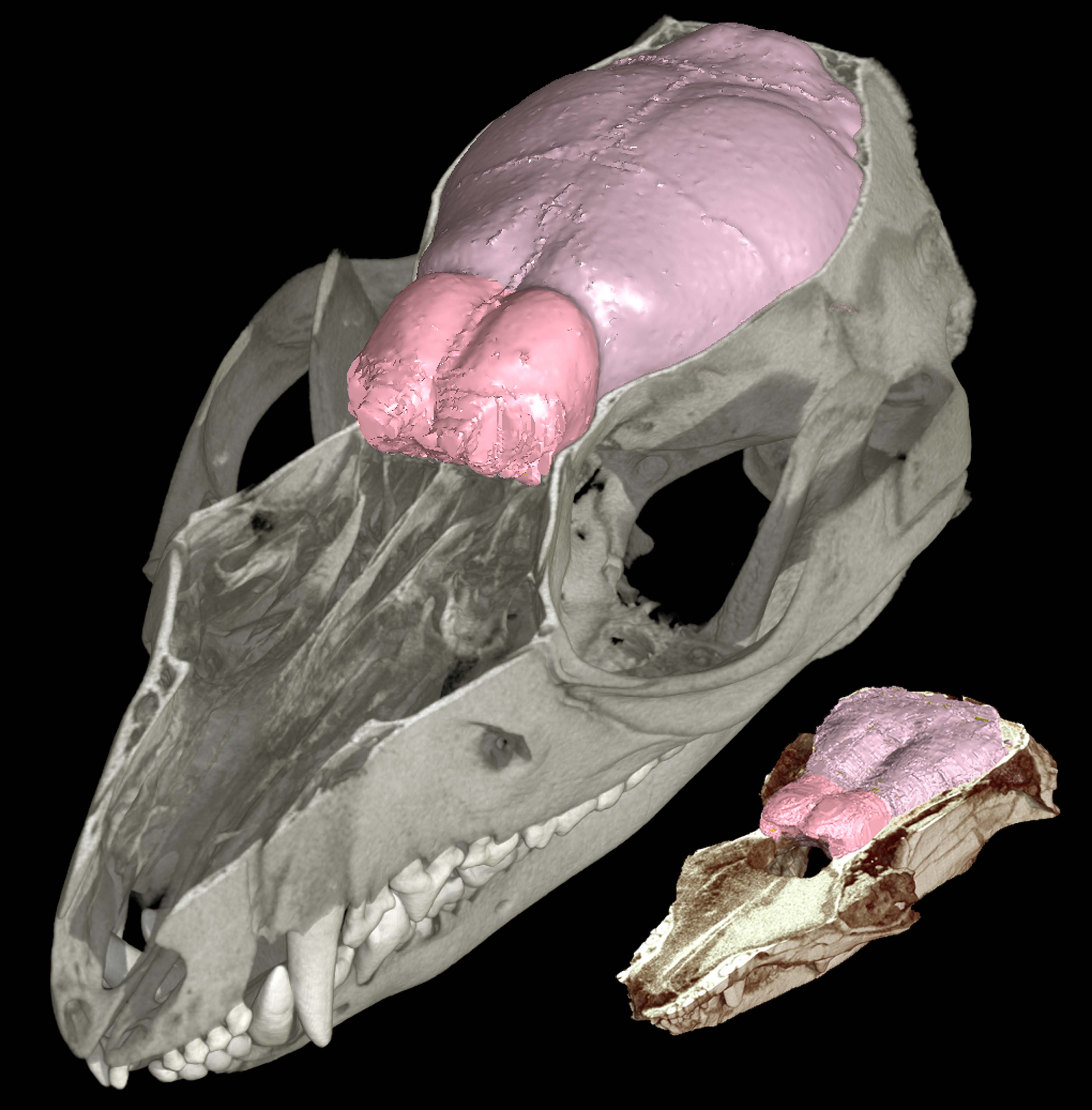
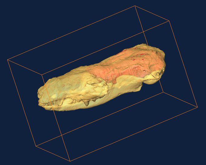
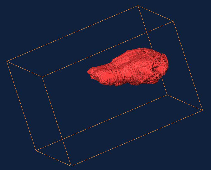
This latest study is the first to use CT technology, similar to medical scanners, to reconstruct the brains of two of the earliest known mammal species, both from the Jurassic fossil beds of China. The 3D scans revealed that even these tiny, 190-million-year-old animals had developed brains larger than expected for specimens of their period, particularly in the brain area for smell.
Among living animals, mammals have the largest brains relative to body size. Scientists have proposed many explanations, but because fossil skulls of early mammals are extremely rare, have been reluctant to cut them open for closer study, thus destroying the fossils. Scientists have mostly relied on comparative studies of living mammals.
“We studied the outside features of these fossils for years,” said Tim Rowe, professor in the Jackson School of Geosciences and director of the Vertebrate Paleontology Laboratory at The University of Texas at Austin, and lead author of the new study. “But until now, studying the brains meant destroying the fossils. With CT technology, we can have our cake and eat it, too.”
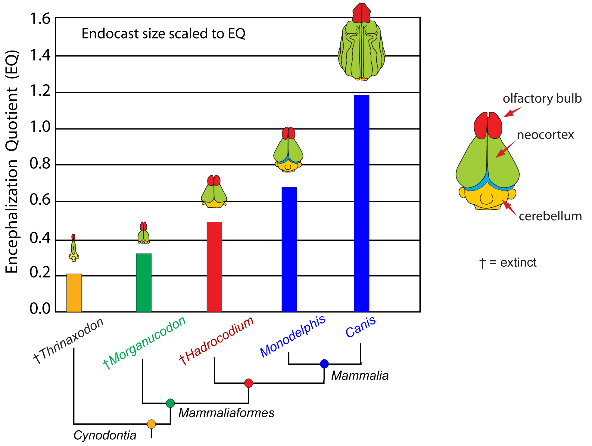
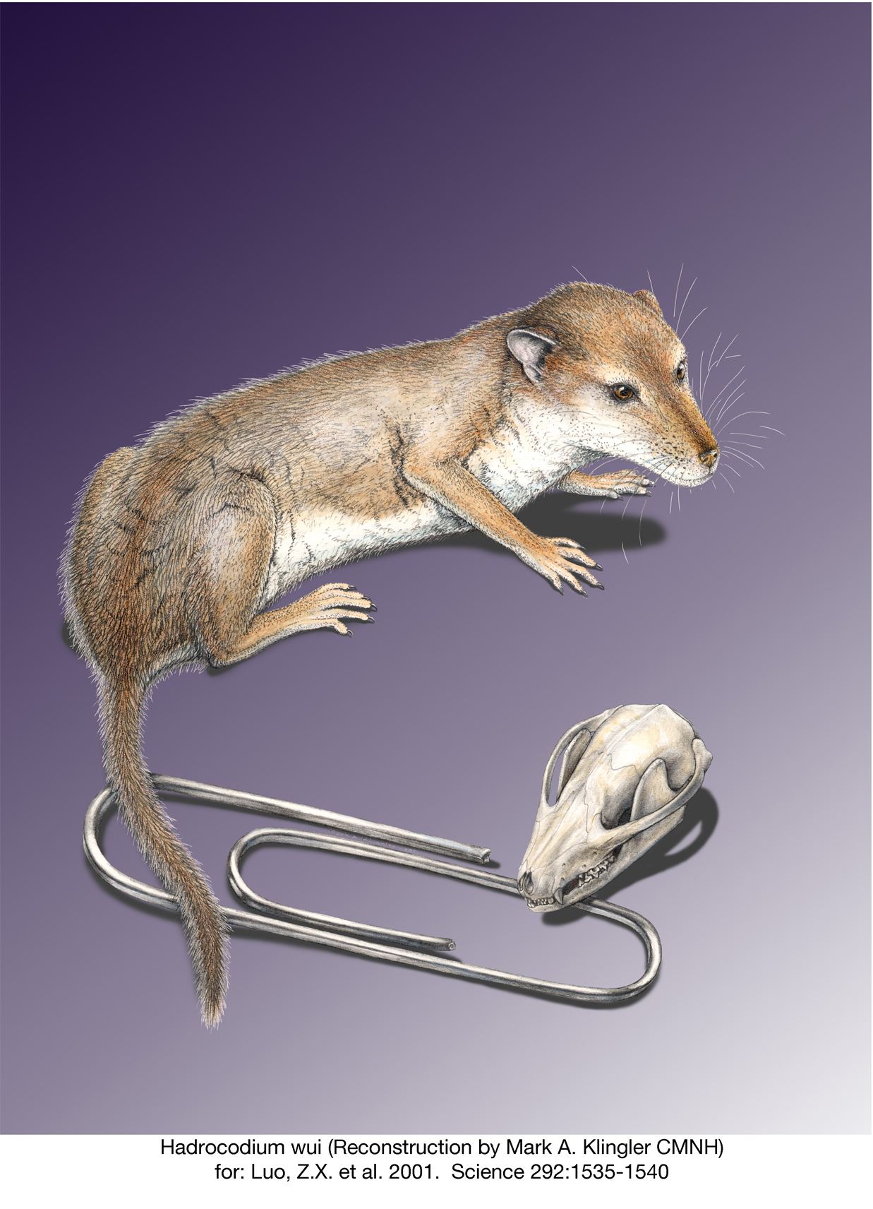
According to the study, other factors leading to larger brains in early mammals included greater tactile sensitivity and enhanced motor coordination. Fossils of some of the earliest mammals, such as Hadrocodium, bore full coats of fur, explaining the need for enhanced tactile sensitivity.

Rowe’s co-authors are Thomas E. Macrini, assistant professor of biological sciences at St. Mary’s University in San Antonio, and Zhe-Xi Luo, curator and associate director for research and collections at the Carnegie Museum of Natural History.
Macrini conducted much of this research for his doctoral dissertation at The University of Texas at Austin, in which he scanned the heads of numerous fossil and living species to visualize the size and shape of their brains.
“This is the most comprehensive study yet undertaken using computed tomography to study the evolution of the mammalian skull,” said Macrini. “And it is exciting to see these new insights emerging from years of intense labor.”
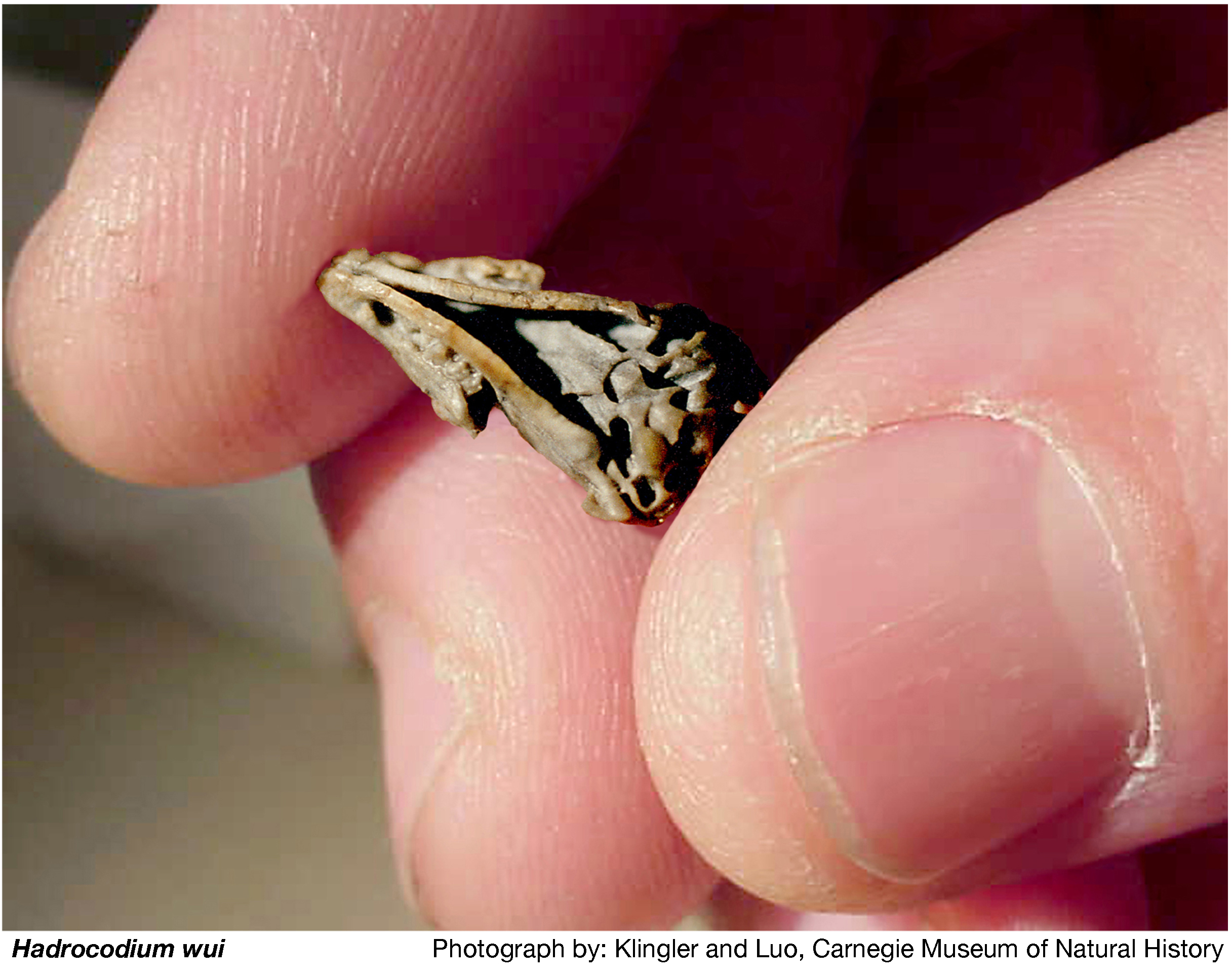
Luo was involved in the discovery and research on the fossils for this study. When he first described the paper clip-sized mammal Hadrocodium ten years ago, he named it for its relatively large cranium despite its appearance so early in the mammalian lineage (“hadro” means “fullness” in Latin and “codium” means “head”).
“I have spent years studying these fossils, but until they were scanned it was impossible to see the internal details,” said Luo. “I was absolutely thrilled to see what the brains of our 190-million-year old relatives were like.”
For this study, the team CT scanned more than a dozen early fossil mammals and more than 200 living species over the past 10 years at the High-Resolution X-ray Computed Tomography Facility at The University of Texas at Austin, a facility supported by the National Science Foundation for researchers around the world. The scans, including interactive 3D fly-throughs, are archived online and available to the public along with nearly 1,000 other specimens on the DigiMorph Web site (www.digimorph.org).
More Info
Anyone who lives with a dog knows not all mammals have equally strong senses of smell. The earliest mammals originated around 200 million years ago. They had robust senses of smell. But since then, some mammals, such as lemurs, have retained that strong sense while others, such as whales, have lost it entirely. Most, such as us humans, are somewhere in between. Rowe says a good analogy for the mammalian brain is a computer.
“You can optimize a computer for anything,” said Rowe. “Even with the most powerful computers, you might optimize them for video or audio or 3D graphics. No one computer is optimized for everything you might want to do. It’s like that with mammals. When you make one sense better, it’s usually at the expense of something else.”
Rowe noted that we humans sacrificed some of our sense of smell for better vision and hearing. Bats traded some of their sense of smell for echolocation; whales for better hearing; and birds for better vision.
“The olfactory system is the single largest set of genes in our genome,” said Rowe. “Like all mammals, we have about 1,000 genes for smelling. But in our case, fewer than half are turned on.”
For more information on the Jackson School, contact J.B. Bird at jbird@jsg.utexas.edu, 512-232-9623.
Images for Reporters (click to download high resolution versions):
|
|
|
|
|
|
|
|
|