A new fossil photography technique developed at the Jackson School of Geosciences is revealing scales, hair and other soft tissues, as well as signs of repair.
BY MONICA KORTSHA
 Enter any natural history museum around the country, and it probably won’t be long until you encounter a cache of fossils — almost certainly big dinosaurs — posed and lit so they look nearly lifelike, not a single tooth or tailbone out of place.
These specimens are meant to inspire wonder, to light a spark that can lead to or strengthen a lifelong interest in science. But the truth is that fossilization is not pretty process. A carcass could have been gnawed upon and scattered by ancient scavengers, bones crushed or distorted by the piling on of millions of years’ worth of rock. And when a skeleton does happen to show up on the surface, weathering and erosion start to whittle everything away.
Making dinosaurs and other fossils look good comes down to the dedication and skill of fossil preparators. However, the preparation process includes techniques that can sometimes lead to scientifically important features, such as soft tissues or fine structures, being unknowingly damaged or stripped away. And sometimes the reconstruction work on fossil specimens is so good that the prepared specimens can fool the very scientists who are studying them. What looks like an actual attribute can turn out to be the result of repair. Biological remains may lurk in places where it looks like nothing is there.
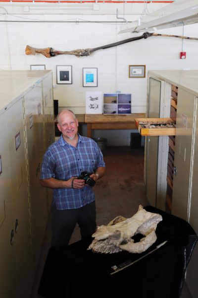
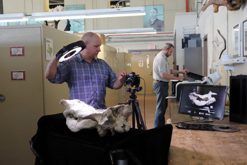
Mike Eklund, a fossil preparator and research associate at the Jackson School of Geosciences, has spent years preparing fossils for scientific study and display. The experience inspired him to take the lead in developing a new photography technique that does not seek to just put fossils in their best light, but under a whole sequence of lights — 17, to be exact — that can illuminate a host of features, including those that would have otherwise remained hidden.
“This is the shortest path to getting the most complete story,” Eklund said.
He calls the technique Progressive Photonics, a nod to the stepwise nature of the approach and its potential for growth as more photographic techniques are incorporated. The approach is starting discussions about how fossils should be researched and prepared. It also has Eklund and others making the case that it’s not just scientists who can benefit from seeing fossils outside of their exhibit-ready looks. Developing the technique has made him an advocate for fossils as not just wow-worthy specimens but as tools to teach how the science of paleontology is done.
MAKE IT PRETTY
When fossils come in from the field, they get shipped off to “beauty school,” Eklund’s term for the preparation process that gets a specimen ready for research or display. At its most basic, the process involves removing enough rock from around the specimen so the fossil can be examined. Depending on the fossil and its envisioned purpose, the preparator then performs a range of interventions. Gluing pieces together, adding missing parts shaped from plaster — or covering sections in protective sealant or even paint in the case of exhibit specimens — are common treatments. Preparation and repair are a necessary part of putting fossils in a state where people can learn from them. The alternative would be paleontologists going cross-eyed trying to mentally piece together drawers of bone fragments, and much less impressive fossil exhibits. But there are many challenges connected to the preparation and repair process. For one, it can be hard to distinguish where rock stops and bone begins. If you scrape away just a bit of bone, any remnants of soft tissue are blasted away with it.
Then there’s the issue of how — or even whether — to distinguish actual fossil from repair materials. That’s a debate that’s been happening practically since paleontology was first practiced as a science in the early 1800s. For example, 19th-century paleontologist Erwin Hinckley Barbour wrote through gritted teeth about how O.C. Marsh, a contemporary of Barbour’s and a famed fossil hunter at Yale University, preferred to display specimens in a coat of black paint — a practice that prioritized looks over substance and forced Marsh to use a damp sponge to distinguish between the porous plaster and mineralized bone. Blacked-out bones have fallen out of vogue now, with most fossil preparators going for a natural look. Nevertheless, the preference for natural-looking specimens combined with advanced repair techniques has caused its own problems. Namely, they can make it difficult to distinguish which fossil features are natural and which are due to getting some work done.
“It’s all being interpreted through the prep process,” said Matthew Brown, the director of museum operations at the Jackson School Museum of Earth History Vertebrate Paleontology Collections. “You take someone and give them hard tools, and they will be shaping the data. People will be looking at the bone, but what you really have is a reconstruction.”
The prepared and repaired fossil affects all research that follows. But the details of what was done to the specimen can be essentially invisible.
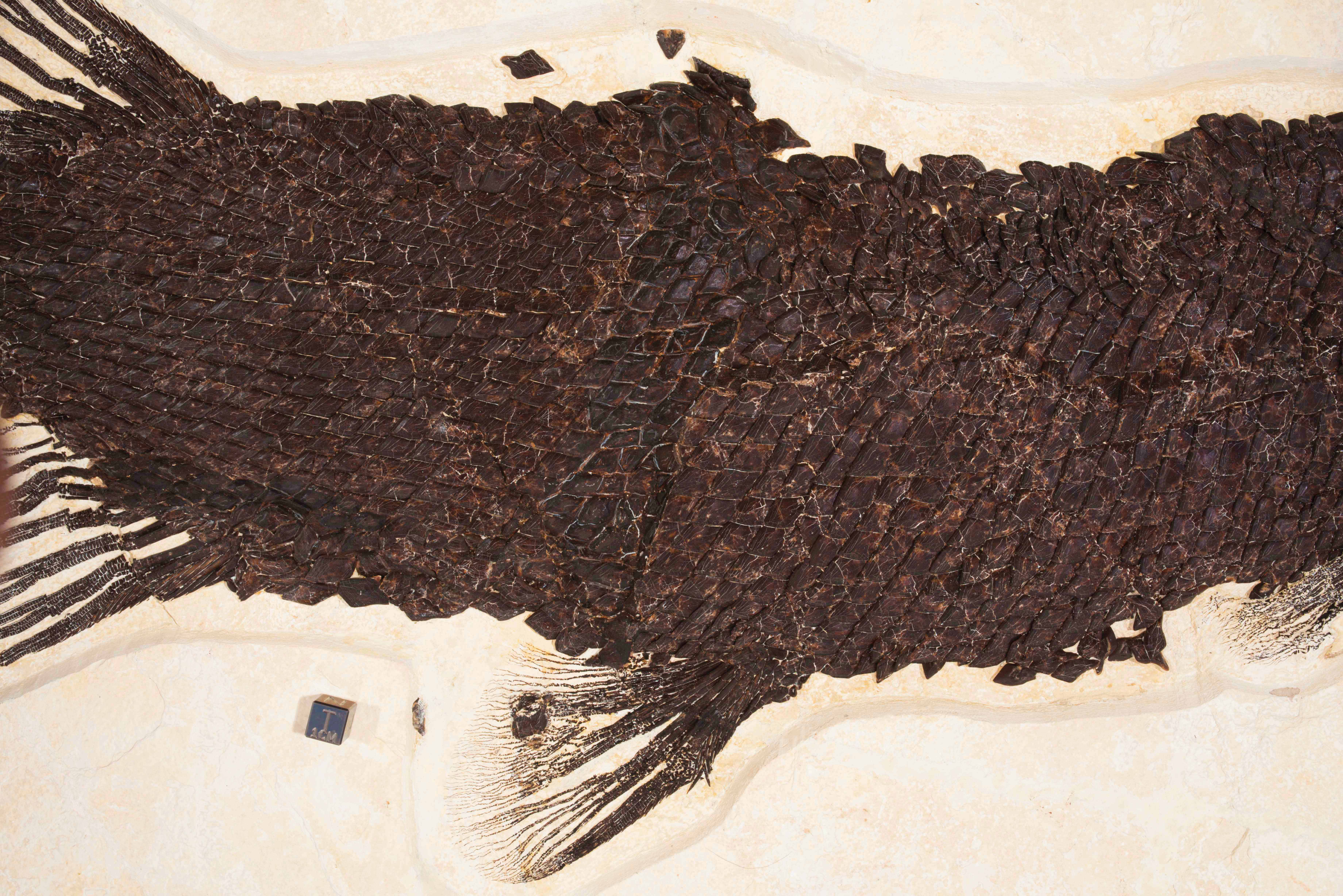
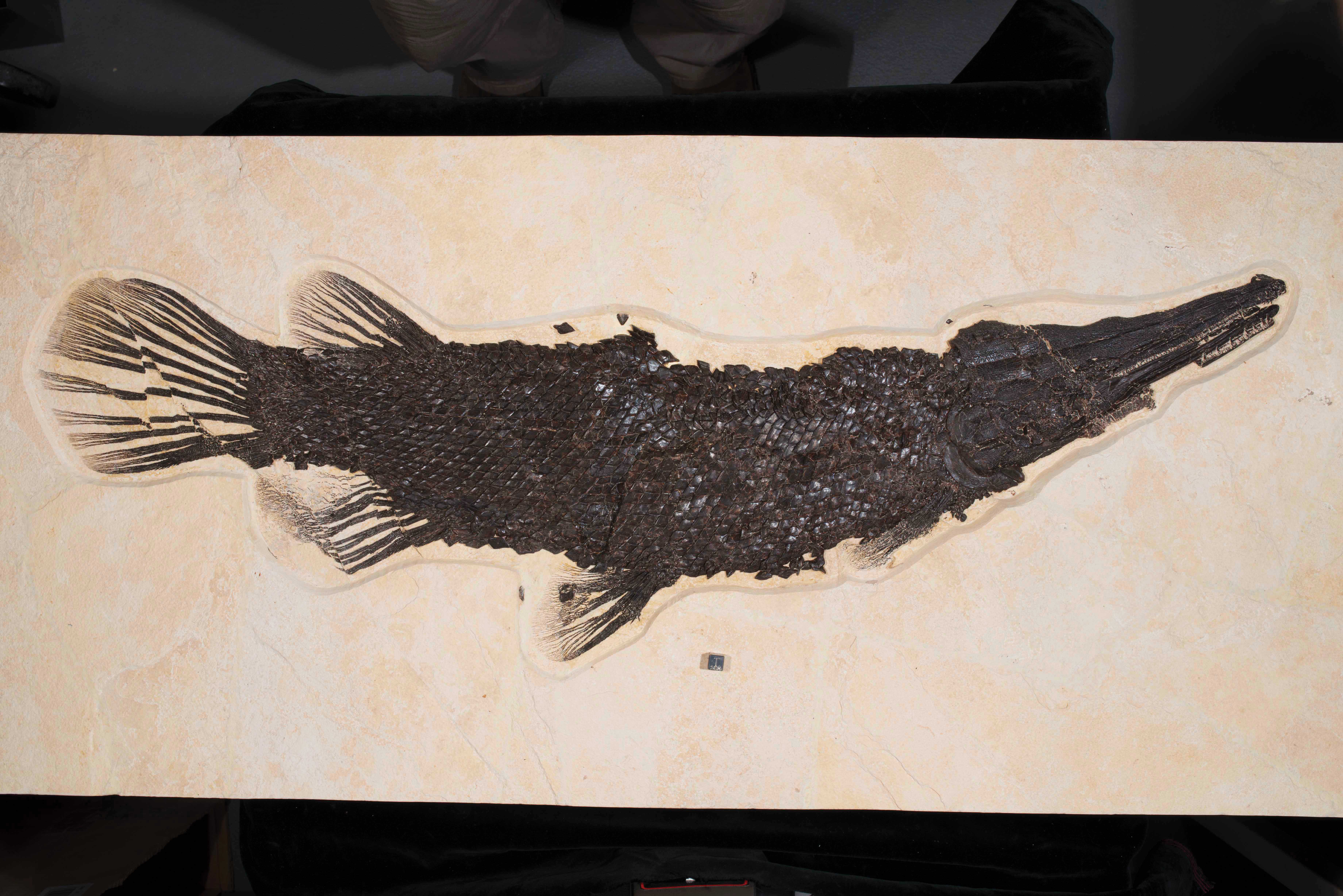
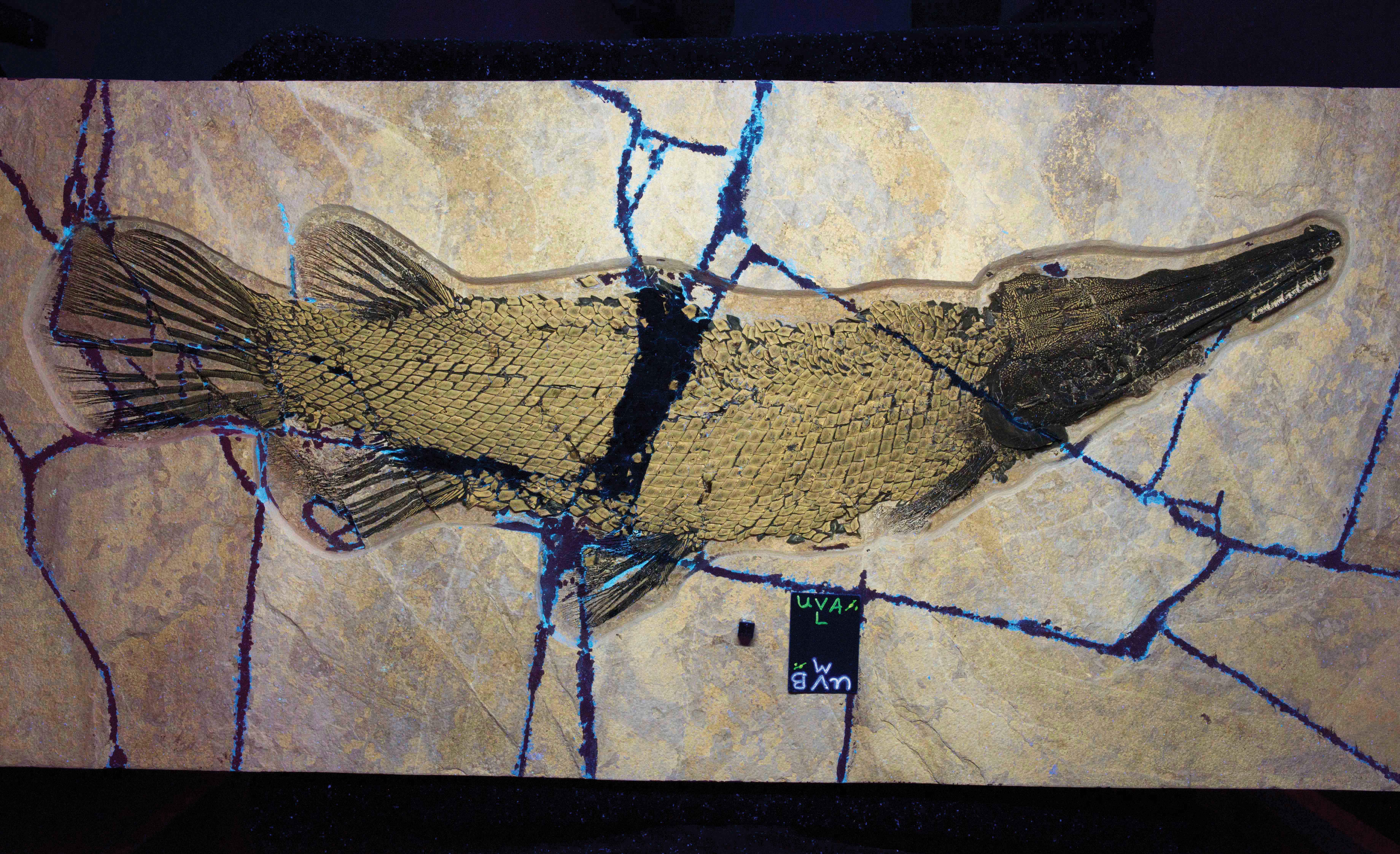
LET THERE BE LIGHT
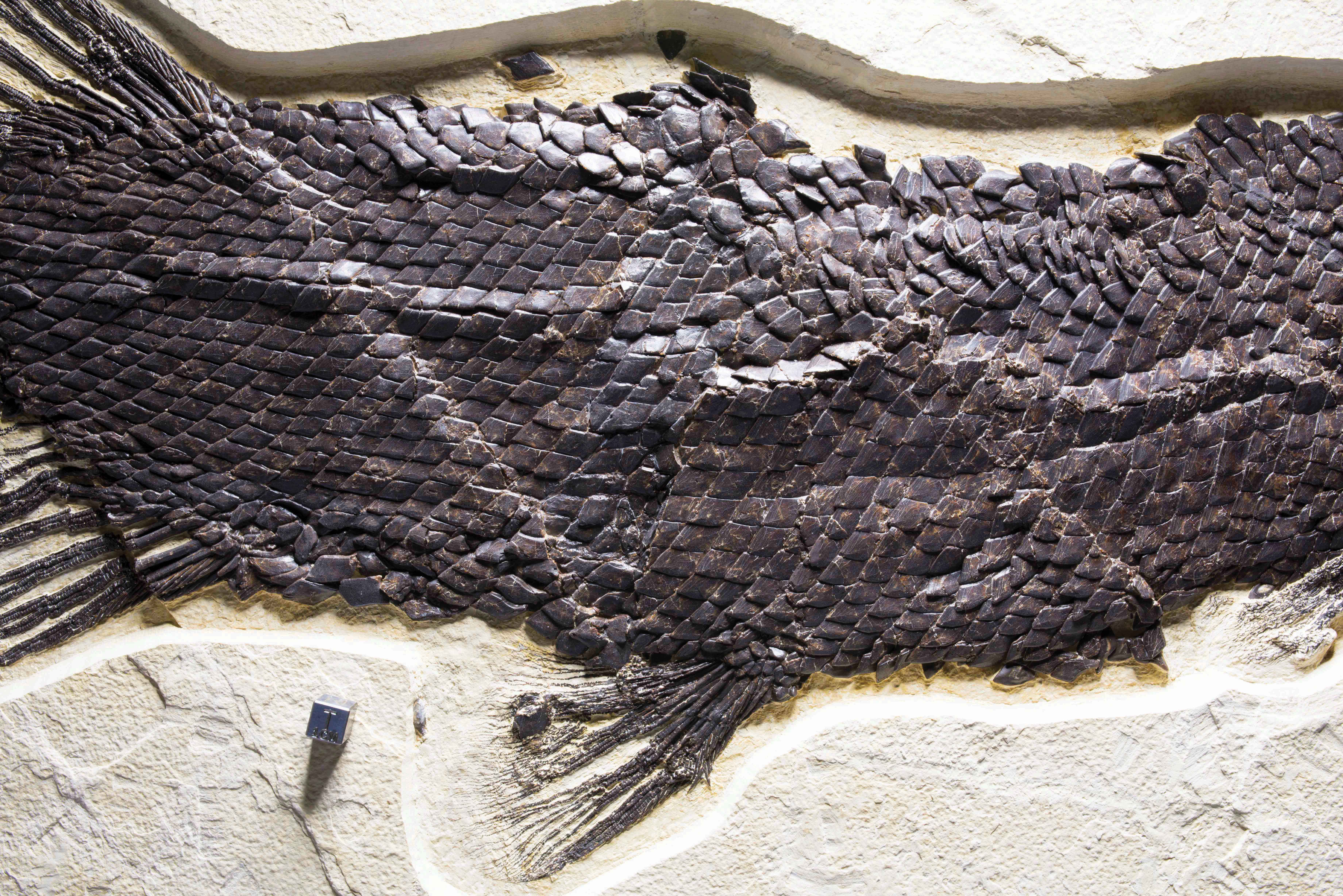
Progressive Photonics can help address the issues inherent to the beauty school process by making different materials in a specimen easier to distinguish. Scientists have experimented with multiple ways of differentiating fossils from their surroundings for decades. O.C. Marsh had his sponge (much to Barbour’s annoyance). And, although illuminating hidden features with UV light has been in use since at least the 1920s, it had never been standardized or even used as a regular practice.
What sets Eklund’s technique apart from others is its meticulous and methodical approach.
He is the first author of a paper published in the Journal of Paleontological Techniques in fall 2018 that serves as a how-to guide for the Progressive Photonics process and provides examples of the technique’s application to various fossils. It covers equipment, safety, and how to shoot the 17-image sequence, which Eklund said takes only about 10 minutes once you have the hang of it. The only thing that changes with each picture is the light source used or the direction of the light.
The resulting photos make it easy for scientists to digitally flip through the images and spot features that appear in
some shots and not others. The photos also serve as a comprehensive digital record of a fossil from multiple vantage points. Uploaded into a collection’s digital repository, the pictures can be a critical research tool for scientists looking to get to know a specimen from afar.
“You’re documenting the history of the specimen while looking for new stuff,” said Jackson School Professor Christopher Bell, who co-authored the paper with Eklund.
Eklund said that he has been developing the Progressive Photonics process for about seven years, and he credits the ongoing support of Brown and others at the Jackson School Museum of Earth History Vertebrate Paleontology Collections with helping get the technique ready for debut to the larger research community.
But even before Eklund got into the paleo world proper, he said that his past jobs — first an accountant, then a custom home builder — put him in the Progressive Photonics mindset by helping him hone an eye for detail and making him a stickler for documentation. So, when
he started volunteering as a fossil preparator at the Field Museum in Chicago — his first preparator gig — he saw how the precision work of preparing and examining fossils could benefit from a standardized documentation process.
However, the technique’s benefits go beyond documentation. The array of lighting types and lighting angles not only helps give a better view than what the naked eye can see, but also helps reveal features that no one expected to find in the first place.
“Fossils are maimed, crushed, distorted,” Eklund said. “We need to look for more than what we think our eyes are seeing.”
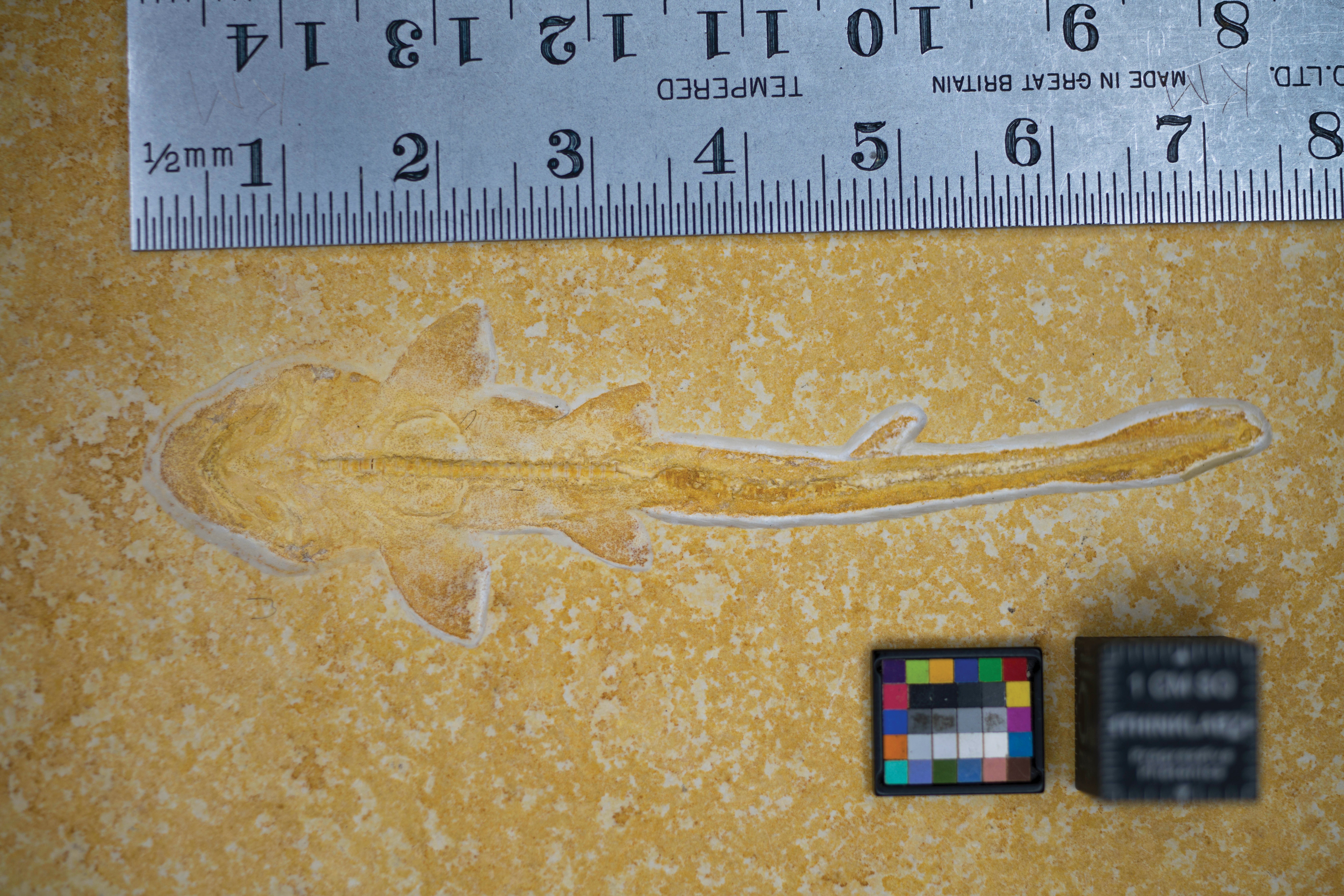
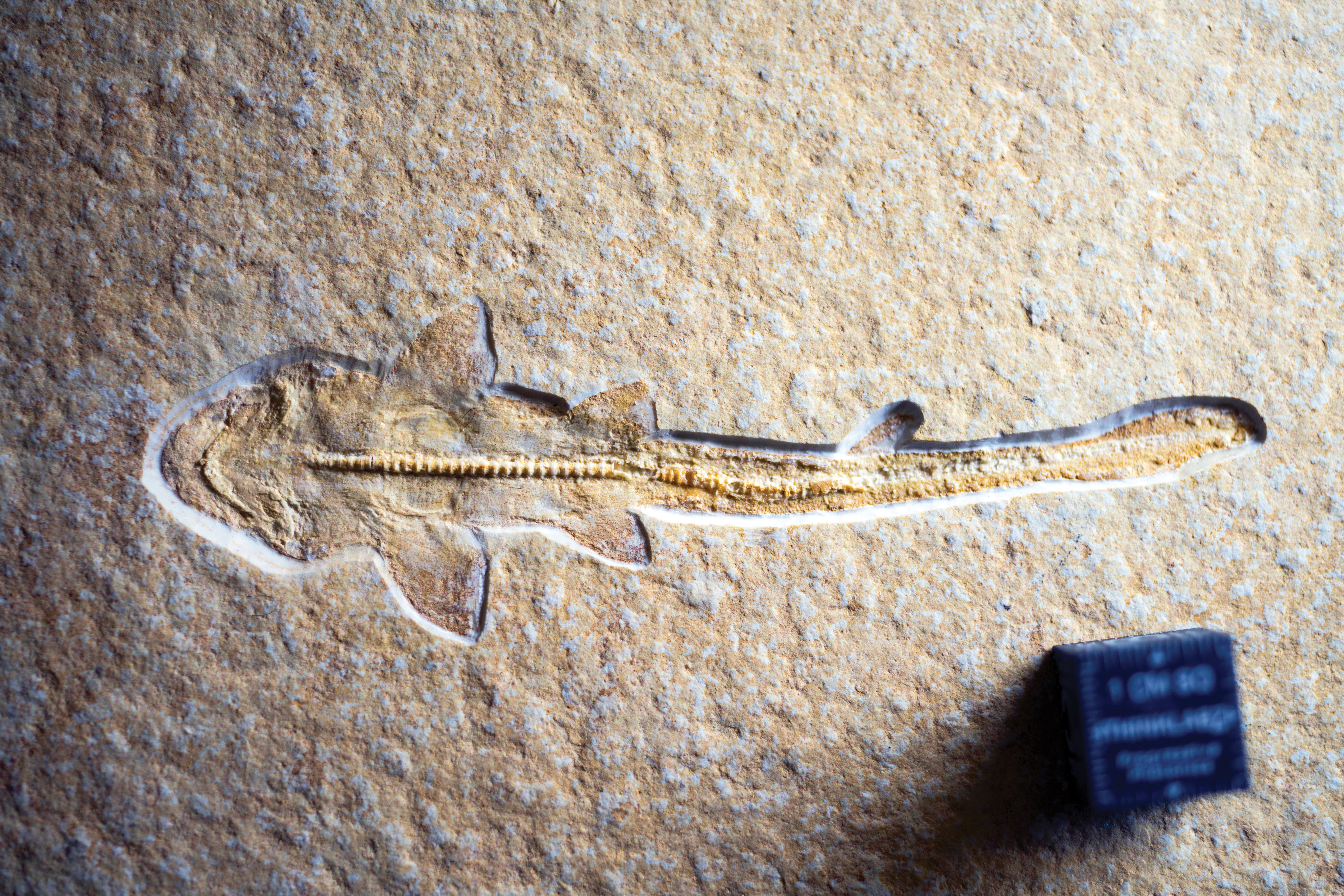
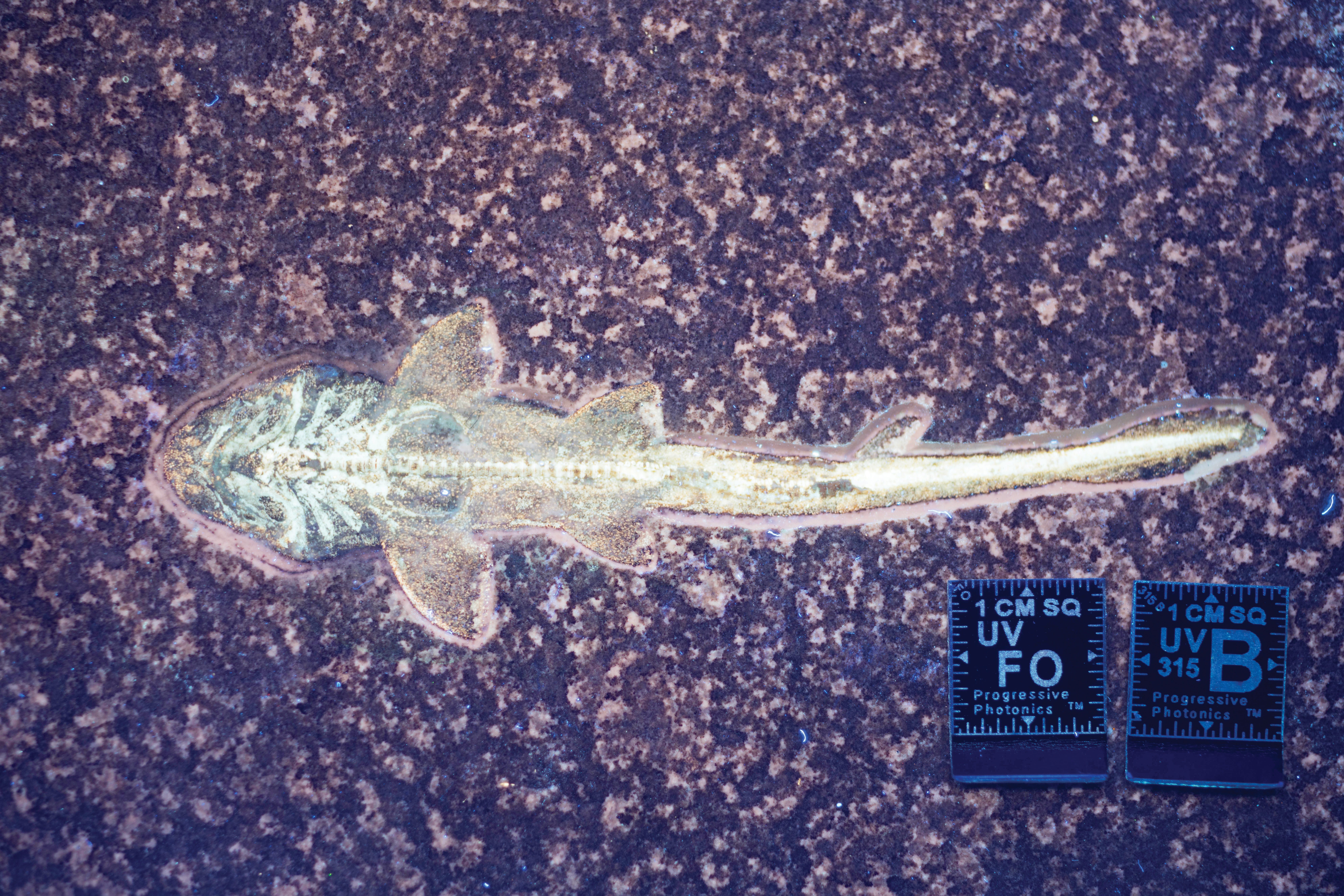
The case of a pinky-finger-size, juvenile shark fossil illustrates this point perfectly. In ambient lighting, the shark appears as a delicate etching. Oblique lighting boosts the contrast, bringing the fossil into high relief. But UV light brings a total surprise: preserved gills, tooth-like scales called denticles, and the remnants of whisker-like sensory organs called barbels. All of these features had gone unseen for decades, blending in perfectly with the surrounding limestone.
Allison Bronson, a lecturer at Humboldt State University, met Eklund and examined the juvenile shark fossil using Progressive Photonics while she was in graduate school at the American Museum of Natural History. She said she usually relies on CT scanning to get a detailed look at specimens. But the technology falls flat with specimens pressed into the surface of a rock slab, which is how delicate animals, such as insects, birds, cephalopods and sharks are frequently preserved.
“There are a lot of things that are flattened that you just can’t scan, and all these things in limestone are like that,” Bronson said. “The best way to look at stuff like that, I think, is Mike’s technique.”
Bronson and Eklund have also examined a different shark specimen While the juvenile shark was lightly stamped in the surrounding rock, this one was still largely trapped within the rock and still in the process of being exposed by preparators. During the Progressive Photonics workup, the shark’s teeth unexpectedly fluoresced under UV light. It’s unknown what exactly caused the teeth to glow, Bronson said. It could be signs of preserved tooth enamel or some other coating; regardless, identifying the glow in the first place could be an important starting point for future research.
Historically, it has been exceedingly rare for scientists to find fossils of vertebrates that contain anything but bones. Internal organs and adornments such as feathers, scales and skin are thin and delicate. Even if they make it through the fossilization process, it’s not a given preparators or researchers will realize they’re there.
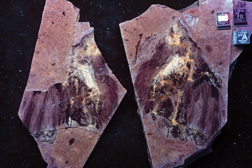
But Eklund said that finding soft tissue during a Progressive Photonics workup is not an unusual occurrence. What scientists previously thought was the rarest of the rare could have just been an oversight.
“This is the next big thing. It’s the next step towards finding things we’ve been missing in the past,” Eklund said about soft tissue finds. “And we know that it’s present way more often than we realized.”
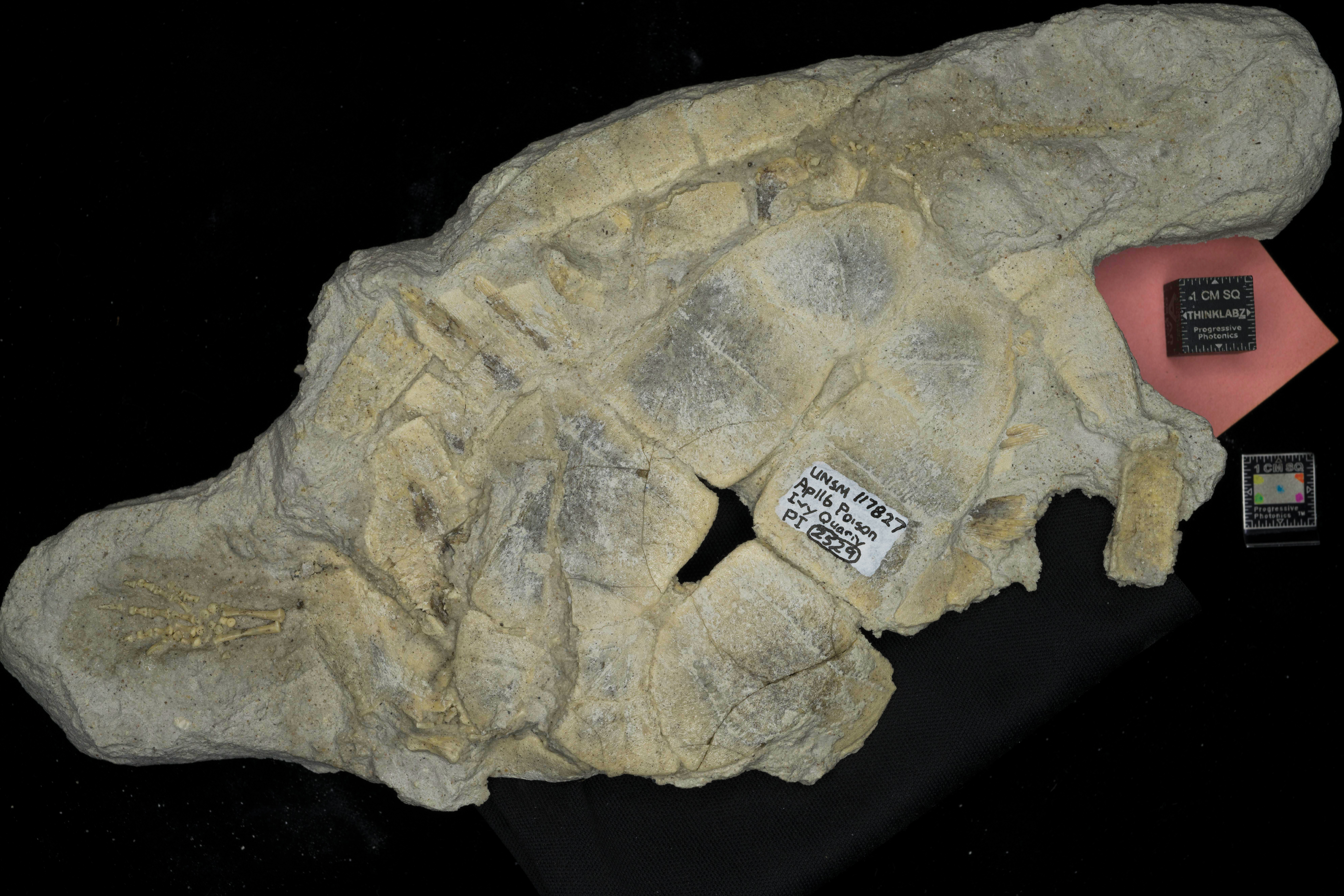
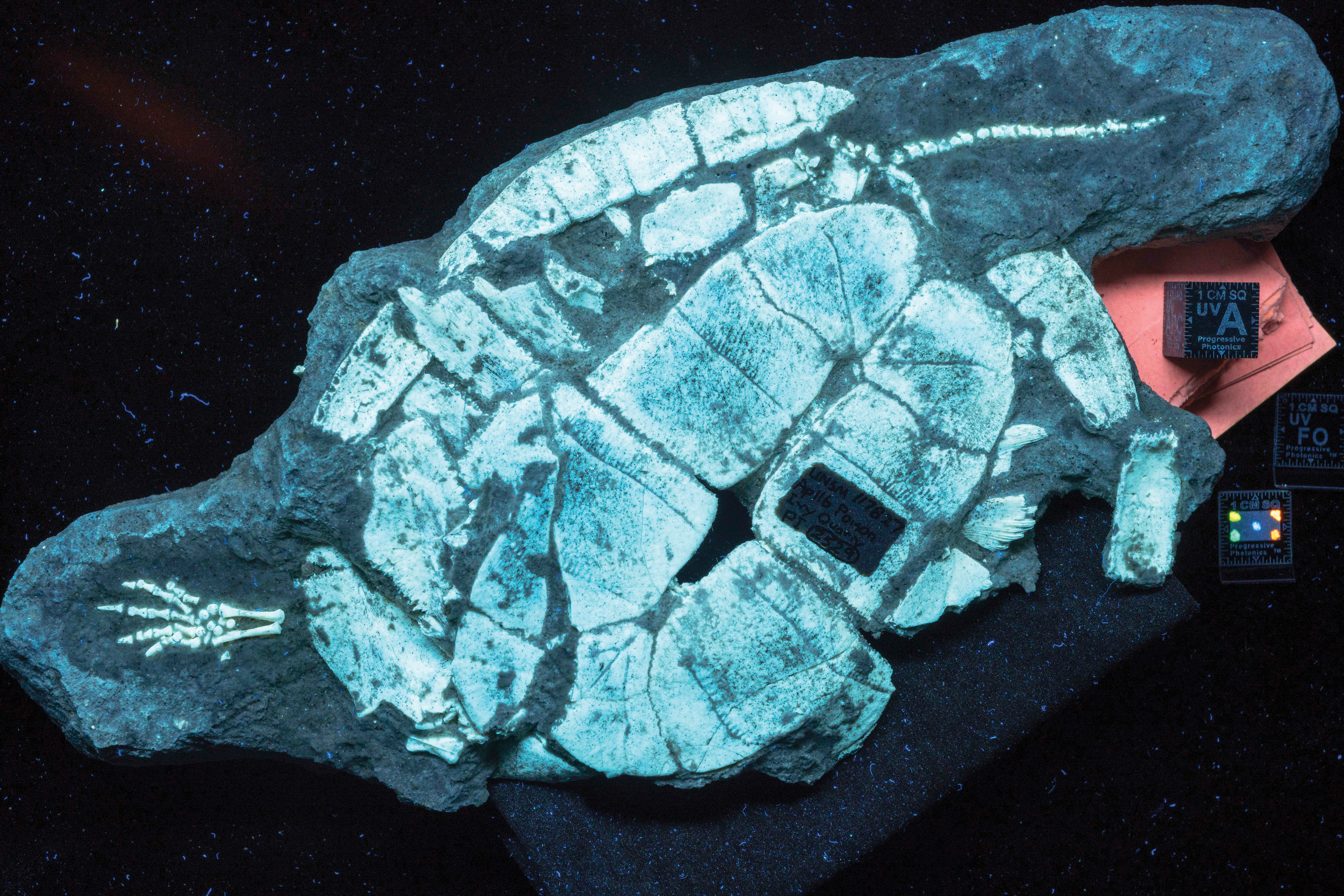
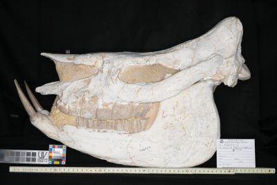
Just as important as the approach’s ability to reveal unseen tissue is the power of Progressive Photonics to show which specimens should not be studied — the ones that are more artistry than anatomy. An Asian rhino skull — or what’s supposed to be one — in the Jackson School’s collections has become a classic example.
It looks like a passable specimen under white light. Shine a UV light on it, though, and the skull is revealed to be a practical mosaic of different materials. Plaster, rhino fossil, and bits of pieces of bone swiped from other unknown fossils all glow with their own distinctive hues.
If a researcher wants to know anything about actual ancient rhinos, they should steer clear of this skull.
For most specimens, though, it’s not an all-or-nothing situation when it comes to research. The same Progressive Photonics session that reveals signs of extensive repair can also distinguish which areas of the fossil are good for scientific study. Eklund gives the example of a pterosaur fossil that was put together from 13 pieces. Certain regions of the fossil were lost to globs of glue. But others gave exceptional insight into the anatomy of the winged reptile. Progressive Photonics offers scientists the insight to unequivocally tell such areas apart.
“You have to think about what you’re looking at,” Eklund said. “And that’s where the suite of images gives you the tools to really digest and interpret what’s present in the specimen. Close proximity is not enough. Likeness is not the real thing.”
Eklund notes that with all the talk of paint, plaster and distinguishing what’s natural from constructed, Progressive Photonics can seem like a ready-made fraud detection tool. But he’s quick to say that’s not the point.
“People look at the system and too quickly jump at associating it with fake, fraud and forgery detection — but those are not relevant terms,” he said. “By having the imaging here I can ask, ‘Is it viable for my study or research purpose? Can I make a statement about the confidence I have in it? Is it acceptable to my research or education needs?’”
In other words, while Progressive Photonics can’t tell you how a fossil came together, it will let you know what’s there. From that point, it’s up to the researcher what to do next, whether that’s write a paper, raise an eyebrow or wave a red flag.
KEEPING IT REAL
Progressive Photonics pulls back the curtain on the preparation history of fossil specimens. That undoubtedly helps scientists analyze their specimens. But Bell said that he thinks the larger public could also benefit from getting to know the work that goes into preparing and displaying fossil specimens. The statuesque skeletons in museums are impressive, but they can give the wrong impression about the science of paleontology: namely, that fossil specimens are much more complete than they actually are.

“That’s a form of deception,” Bell said. “It’s benign deception. It’s not done with the intent to deceive or lie. It’s an intent to have a visually captivating representation of past life on Earth — but you’re ceding the interpretive context.”
Jackson School Professor Christopher bell helped develop the progressive photonics method and co-authored a paper on it with Eklund.And when a display isn’t forthcoming about what’s real and what’s not, it can give fodder to those who want to undermine a scientific world view in general, he said.
In contrast, exhibits that employ the principles of Progressive Photonics — seeking to highlight unseen features, those left behind by nature and those added in museum prep labs alike — could provide more information about the specimen and the scientific reasoning that informed the display process.
“Why not talk about it?” said Bell. “From my perspective, we’re missing out on a really important educational opportunity to talk to people about the confidence we have when reconstructing the past and the places where we do have differing degrees of uncertainty.”
The Smithsonian Institution has embraced this line of thinking. In June 2019, the National Museum of Natural History reopened its hall of fossils after an extensive five-year renovation, the first one since the fossil hall opened in 1911. The fossil displays include casts of bones articulated into lifelike poses — a dog-size Stegoceras dinosaur using its back foot to scratch the top of its domed head, a Tyrannosaurus rex chomping on the frilled crest of a Triceratops, an extinct species of horse rearing up like a bony replica of Silver, the Lone Ranger’s steed. These mounts serve as a reminder of the amazing animals that once walked the Earth. But when it comes to real bones, the distinction is clear. All synthetic materials are painted a shade darker than the surrounding bone, said Steve Jabo, a preparator, researcher and collections assistant in the Smithsonian’s Department of Paleobiology. That wasn’t always the case. Many of the old mounts gave the illusion of a complete specimen by painting plaster the same shade as bone, or even painting over the bone itself.
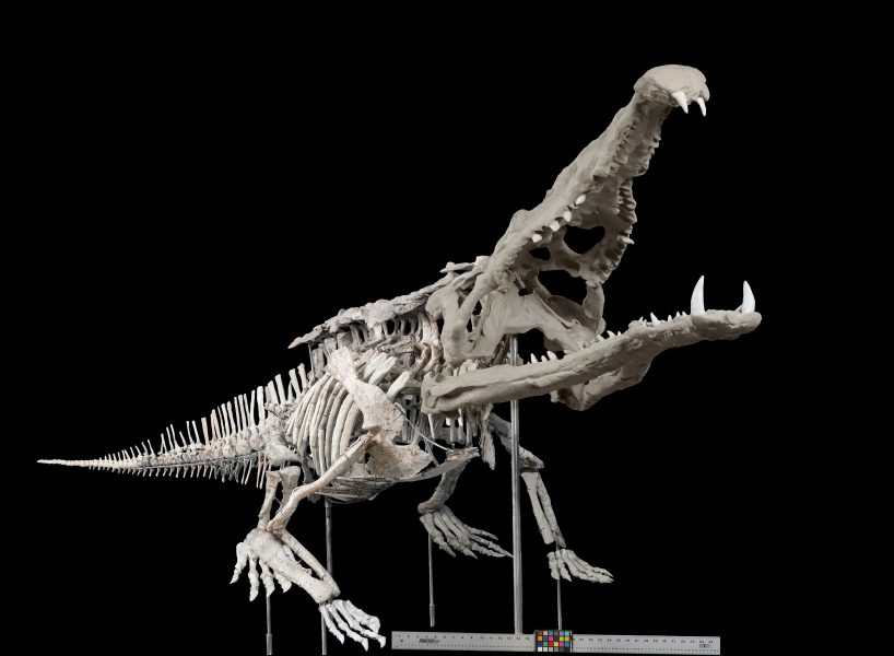
Part of the renovation process involved removing the illusion of a complete specimen when preparators were just working with parts.
“We made the decision to go back and do that uniformly,” Jabo said. “We don’t want to trick people, especially the researchers, the people who are doing active research on our research specimens. Because deciding if something is real or not, sometimes that’s hard to do.”
The new exhibit also features a UV light box. At the push of a button, delicate crab fossils from the famed German Solnhofen Limestone go from blending in with the surrounding rock to popping out in a shade of glow-stick orange. Eklund played a key role in choosing the lightbox specimens, which were selected from among a number of specimens in the Smithsonian collection that got the Progressive Photonics treatment during a research visit.
In addition to helping with the exhibit, Jabo said that seeing Progressive Photonics in action has changed his approach to fossil prep, mainly, to go easy on the consolidant (which can cover small or delicate parts and complicate the chemistry research) and to stop and check for soft tissues.
“It has affected how I do prep,” Jabo said. “In the process of doing things, I can stop, get a light on it to look and see if there’s anything. If something cleaves along the bone, that’s the perfect time to look at it because that’s where you’re going to find any soft tissue preservation.”
The work of fossil preparators sets the stage for a fossil’s future — how it’s interpreted, how it’s displayed, what is seen and what goes overlooked. But just like the many hidden fossil features that Progressive Photonics has helped bring to light, the critical role of preparators in paleontological research usually remains in the shadows.
Preparators are frequently volunteers. They come from a variety of backgrounds and don’t always work within the usual research spheres, said Brown, whose own career in paleontology started when he was a volunteer fossil preparator while in high school.
“It’s a quirk of paleo; the person who discovers a fossil is usually not the preparator,” Brown said. “The preparator
is the hidden hand.”
Progressive Photonics helps reveal the impact of that “hidden hand” and other features that would have otherwise remained hidden to the human eye. By spreading the word and practice of the technique to preparators and scientists alike, Eklund said that he hopes to remove some of the uncertainty that’s inherent to studying fossils. You might not know much about how a fossilized animal lived, but you can at least know whether the fossil is a good starting point for asking questions, Bell said.
“When you go back and image these fossils, you can now say ‘this part’s paint, this part’s plaster and now we know,’” said Bell. “We don’t have to worry. We don’t have to guess.”
The University of Texas at Austin
Web Privacy | Web Accessibility Policy | Adobe Reader


