JEOL 6490 Low-Vacuum Scanning Electron Microscope
Location
JGB5.108
Overview
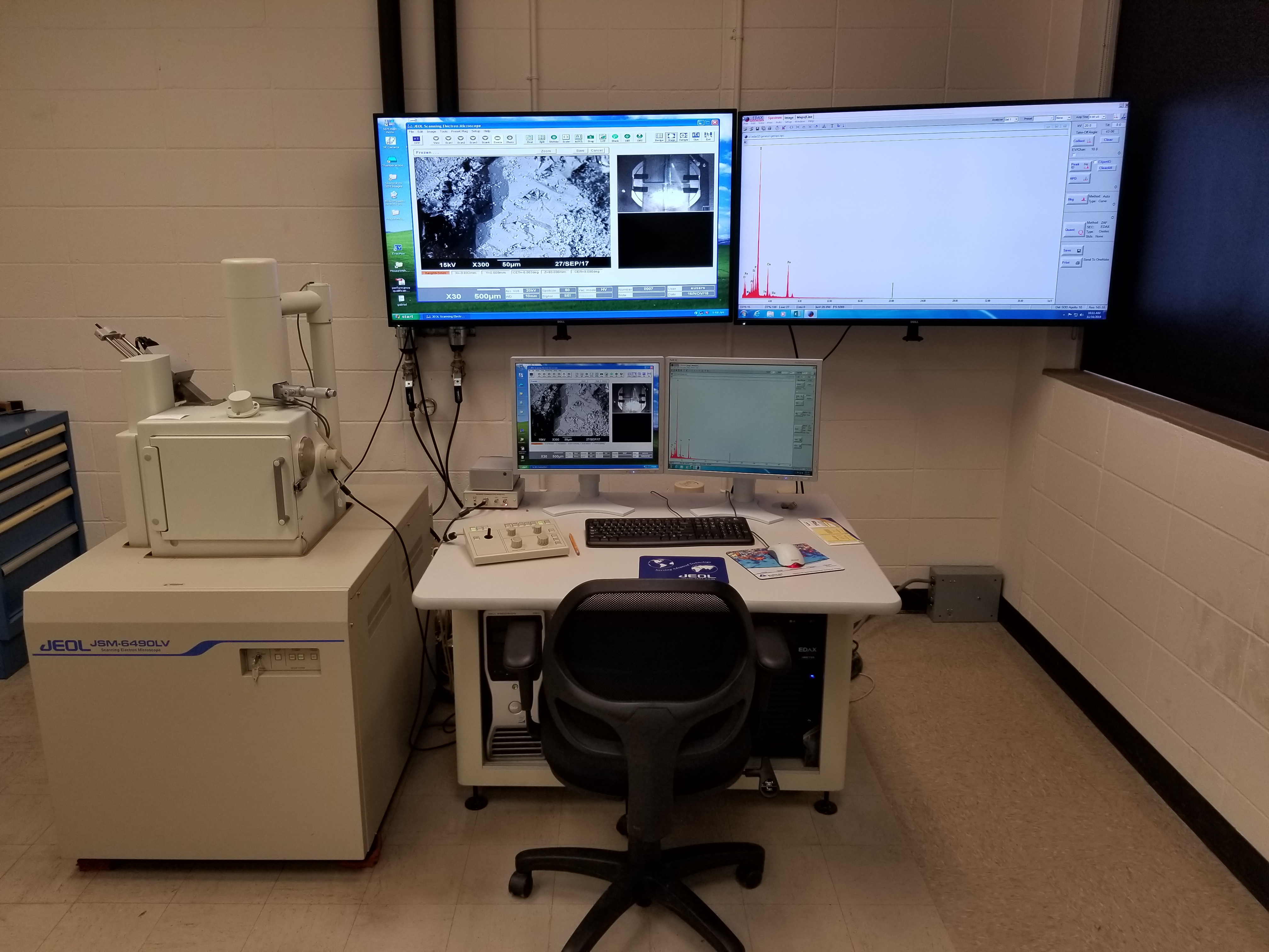
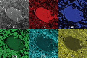
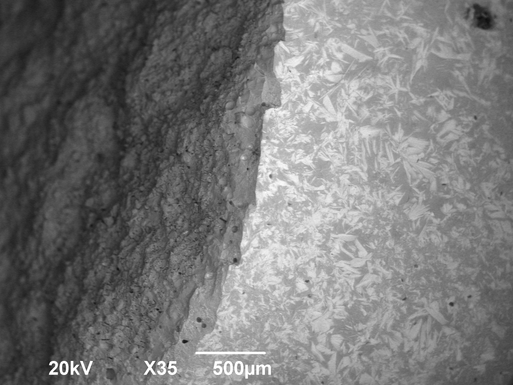
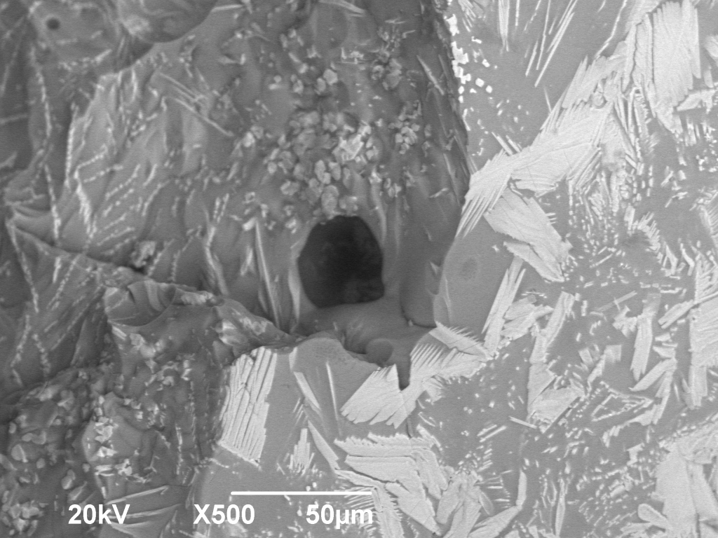
Installed in 2008, our W-filament JEOL 6490LV SEM has secondary electron (SE), back-scattered electron (BSE), and energy-dispersive X-ray (EDS) detectors. The 6490 can operate in high- or low-vacuum modes, allowing sub-micron-scale imaging and X-ray compositional data collection without needing to coat or modify your sample. The EDS detector can be used to create a qualitative and semi-quantitative elemental analysis of sub-micron point volumes, X-ray mapping over mm-scale single fields of view, and X-ray line scans.
This SEM received a major upgrade. We installed a Gatan PanaCL detector, allowing the collection of cathodoluminescence signals; an Oxford Instruments Nordlys Nano EBSD detector; and upgraded the EDS detector to the far more powerful Oxford Instruments X-Max 50. The EBSD and EDS detectors will be configured for simultaneous collection, and both will run through Oxford Instrument’s very user-friendly Aztec software. After this upgrade, fully automated Large Area Mapping is an option for the collection of SE, BSE, EBSD, EDS, and CL data.
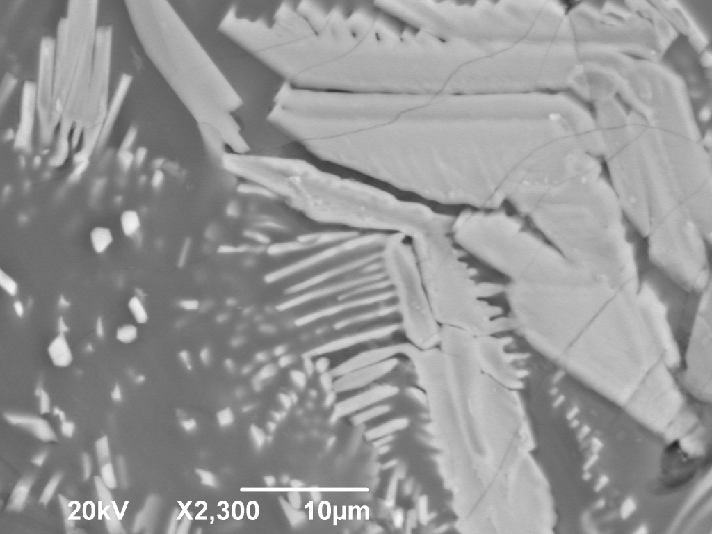
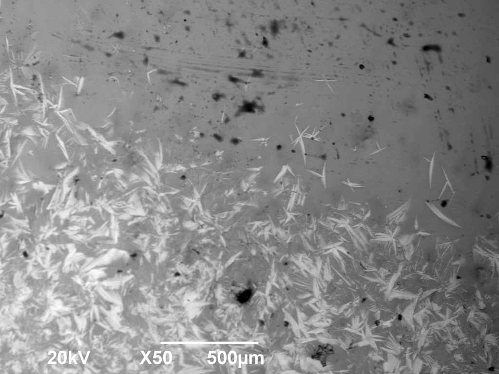
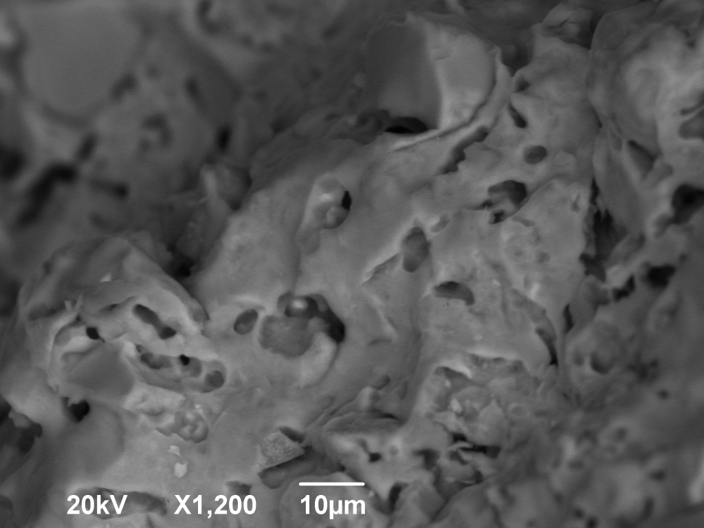
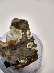
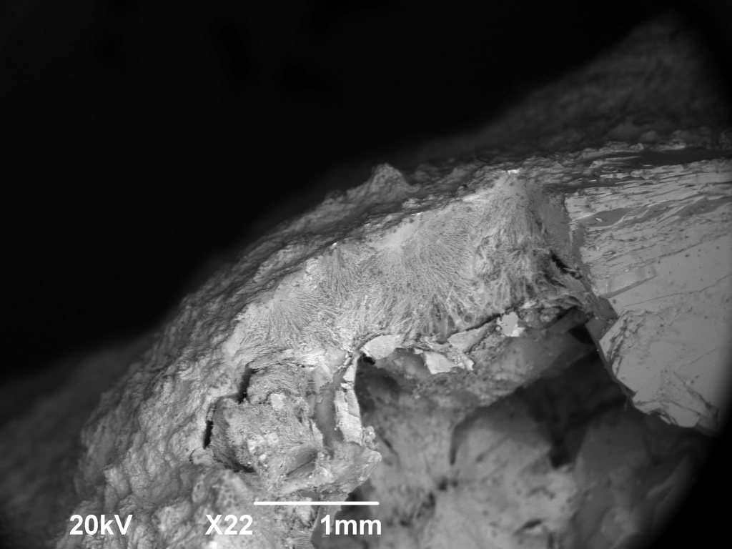
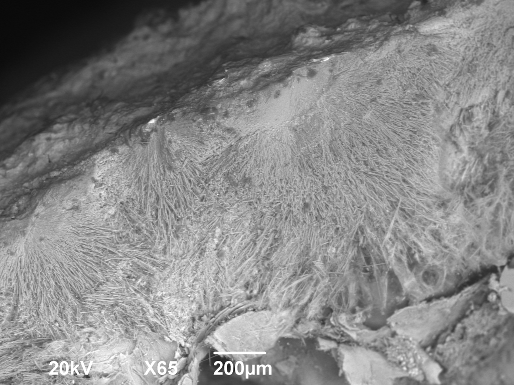
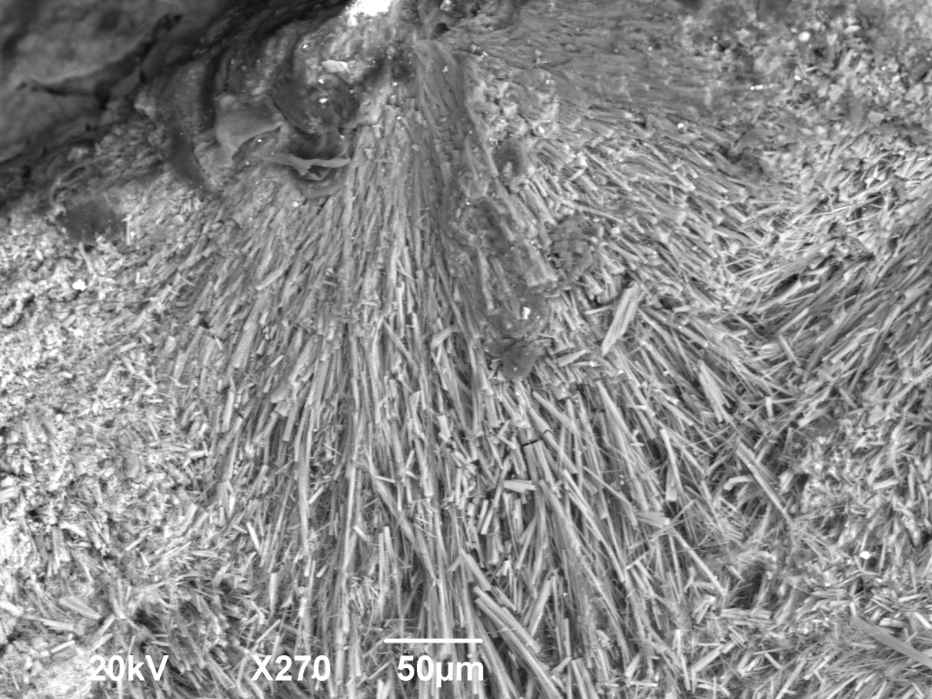
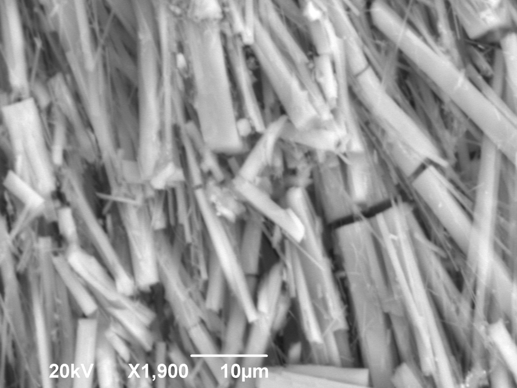
Examples
Requirements of Samples
- Because the SEM can operate in high- or low-vacuum modes, a conductive coating is not required for insulating samples. However, conductive samples (or those with a conductive coating) are preferable for optimal image acquisition and chemical analysis. Carbon coating is available in the DGS Electron Microbeam Laboratory.
- Samples must be dry (water is the main concern, but some oil-saturated samples might also be unsuitable).
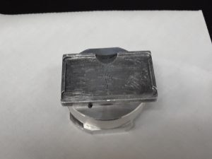
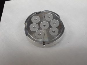
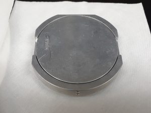
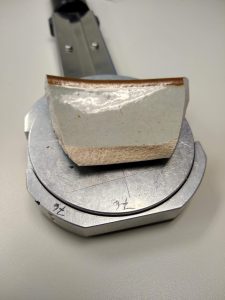
Sample Shape and Size
- Typical samples are thin sections and materials < 1 cm in diameter.
- Four 1-cm diameter samples can be loaded into the SEM simultaneously.
- Samples up to 5 cm in diameter may also be analyzed, with the warning that large samples pose the possibility of colliding with the detectors.
- The stage can be tilted up to 55° from the horizontal.