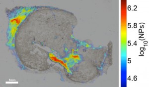
Figure. Tissue histology qPA image of human squamous cell carcinoma overlain by bright-field optical image of iron-oxide nanoparticles.
Jason Cook, Wolfgang Frey, and Stanislav Emelianov
UT Austin Dept. of Biomedical Engineering
We designed a custom photoacoustic microscope with optical resolution to image and quantify nanoparticles in cells and thin tissue slices. Quantitative photoacoustic (qPA) imaging is based on the dependence of the photoacoustic signal with both the nanoparticle quantity and the laser fluence. Using multiple fluences, the effects of optical scattering were eliminated to enable accurate quantitation in turbid environments. To assess the accuracy of qPA imaging, nanoparticle-loaded macrophage cells were quantified and compared to ICP-MS. After validating the accuracy, qPA imaging was then used to image unstained histology slides from a nanoparticle-loaded xenograft human squamous cell carcinoma (Figure). Our results suggest that qPA imaging may be used in many biological nanoparticle applications including molecular targeting studies in vitro and in vivo, validation of clinical molecular imaging, and biodistribution studies for the assessment of toxicity.
© 2025 Jackson School of Geosciences, The University of Texas at Austin

