Kane Caves Images
Click on photographs to see the full image. Photos were taken by A.S. Engel (aengel@geol.lsu.edu) (unless indicated otherwise) and permission needs to be requested to reproduce these images.
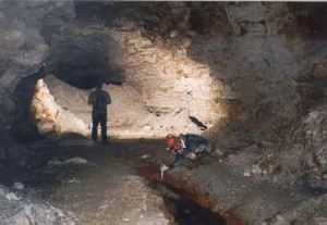
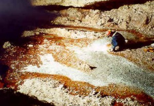
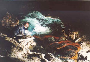
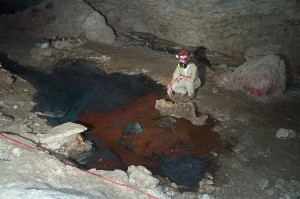
Lower Kane Cave, May 1999. This is the Iron Pool area described by Egemeier (1981
Kane Cave, August 2000. The same Iron Pool area, with up to approximately 5-10 cm of water covering the cave floor
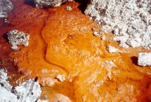
Extremely thick orange-red microbial mats near Lower Spring, August 2000. Pen is near lower left corner for scale.
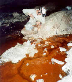
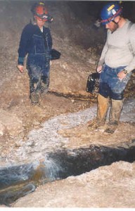
Dr. Libby Stern sampling water flowing through the mats.
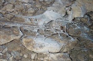
White microbial mats forming a dense filamentous plume from the Upper Spring, August 2000
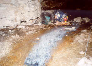
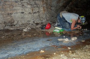
Scott Engel and Megan Porter searching for aquatic and terrestrial invertebrates near white filamentous mats above the Upper Spring and Iron Pool area, August 2000.
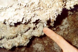
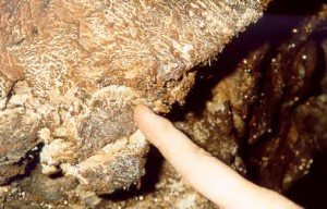
Gypsum paste (or moonmilk) and gypsum needles with biofilms. The pH of the paste and drops was measured to be 0, as well.
Left: Lower Spring oriface and white filamentous mats, May 1999.
Upper Spring pool and oriface with black and red mats, May 1999.
Megan Porter sampling Fissure Spring mats. August 2002.
Fissure Spring white filaments and gas bubbles (methane), August 2002..
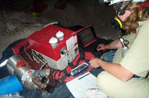
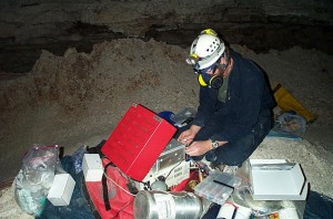
Katrina Mabin running samples on the GC – in the cave! Do the data dance!!!
Witnessing history of field science!!!Phil Bennett setting up gas chromatograph in cave – first time ever! A very exciting moment… August 2002.
Katrina Mabin on the edge of the Upper Spring pool. August 2002.
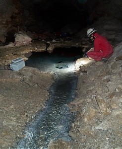
Libby Stern at the outflow of Upper Spring pool, August 2002..
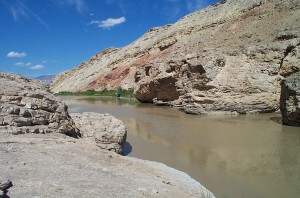
River canyon, looking north. August 2002..
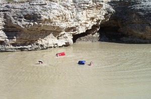
Melissa Edwards, Katrina Mabin, and John Deans swimming in the Bighorn River after sampling Hellspont Cave. oooo fun! August 2002.


