Facilities
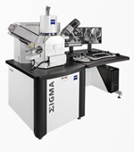
SEM CL Structural Diagenesis FacilityOur main scanning electron microscope and CL detector are designed to collect high-resolution structural diagenesis information over wide areas of rock. The Zeiss Sigma High Vacuum Field Emission SEM with an Oxford X-Max 50 Silicon drift detector (SDD) and a Gatan MonoCL4 system is specifically configured for large-area SE, BSE, EDS, and CL imaging of geologic samples. The Zeiss Sigma HV FE SEM is equipped with a GEMINI column that is designed to work at low Kv in order to minimize sample damage. The large chamber with a 5-axes cartesian stage is capable of covering a 5’’ x 5’’ travel distance for large area mapping. The BSE detector is mounted on the pole piece allowing for rapid transition from SE to BSE imaging. The Oxford X-MAX has a SDD size of 50 mm2 providing a superb resolution and low energy detectability. This instrument also has two independent modes for CL imaging: The MonoCL4 system provides crisp monochromatic, panchromatic, and colored-CL images with high spatial and spectral resolution. The Zeiss VPSE detector allows for CL imaging at low magnification. This VPSE detector is enhanced with an Indigo CL detector to facilitate imaging of specimens containing carbonates without artifacts. Aztec and Digiscan software allow for automated imaging and stitching of large areas. More information about this facility Lab contact: Esti Ukar Recent news: The new device is fully operational, September 2014. |
|
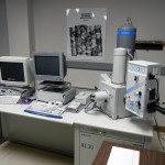
SEM with CL mosaic capabilityOur venerable Phillips XL30 scanning electron microscope equipped with an Oxford Instruments MonoCL cathodoluminescence system for CL mosaic and fracture population inventory analysis was retired in 2014. The Phillips XL30 SEM was operated at 12–15 kV and at large sample currents for panchromatic CL imaging. Color CL images were obtained by combining three greyscale images produced using red, green, and blue filters. We estimate that this instrument imaged more than 50,000 microfractures during it’s SDI campaigns. It was also the instrument used to first uncover nanoporosity in shale. Lab contact: Steve Laubach Recent paper: Hooker, J.N., Laubach, S.E., and Marrett, R., 2014, A universal power-law scaling exponent for fracture apertures in sandstone. Geological Society of America Bulletin 126, 9-10, 1340-1362. doi: 10.1130/B30945.1 | view at GSW |
|
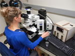
Fluid Inclusion LaboratoryOur Fluid Inclusion Laboratory includes a FLUID, Inc.-adapted USGS-type gas flow heating/cooling stage and a Linkam THMSG 600°C programmable heating/cooling stage. The stages are used on a Olympus BX 51 optical microscope, fitted for use with transmitted, reflected, and UV light, and equipped with 4×, 10×, 40×, 50×, and 100× objectives and 15× oculars. The microscope is also equipped with a 5.0 Megapixel OLYMPUS Q-Color5TM digital camera coupled with QCapture PRO digital imaging software. The lab is fully equipped with sample preparatory facilities for cutting and preparing doubly polished thin and thick sections. Lab contact: Andras Fall Recent paper: Fall, A., Eichhubl, P., Bodnar, R. J., Laubach, S. E., Davis, J. S., 2015, Natural hydraulic fracturing of tight-gas sandstone reservoirs, Piceance Basin, Colorado. Geological Society of America Bulletin, 127, 1-2, 61-75. | view at publisher |
|
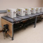
Hydrothermal Experimental FacilitySDI’s hydrothermal experimental system consists of six externally heated cold-seal pressure vessels with operation temperature and pressure conditions of up to 800° and 350 MPa. A key element of current SDI research is to perform laboratory experiments on quartz cement growth in artificial fractures to explore the interaction between fracture opening and fracture cement precipitation, growth interactions between host rock and mineral cement, and how this interaction affects patterns of fracture aperture and cement textures. The experimental observations provide calibration for our diagenetic and geomechanical numerical model efforts, guidance for interpreting fracture cement patterns such as crack-seal and euhedral cements, and constraints on rates and durations of fracture cementation. Lab contact: Andras Fall Recent news: Inaugural experimental results were successfully obtained in Spring 2014. An experimental campaign is currently underway. |
|
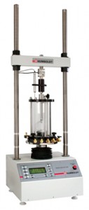
Geomechanics Testing Laboratory at PGE & DGSThe Geomechanics Testing Laboratory has capabilities for a wide range of rock property tests and experiments. The lab houses several load frames, which provide for the deformation of specimens over a range in size, geometry and load. Tests in the Humboldt Manufacturing Company’s hm – 3000 Digital Loader include the Semicircular Bend Test, the Brazilian Test, and Unconfined Strength tests. Addition of a Teledyne ISCO syringe pump provides servo control of confining pressure for measurement of triaxial behavior (Young’s Modulus and Poisson’s ratio). Measurements of deformations and failure in small scale 3- and 4- point bending apparatus can be used to determine Young’s Modulus, Fracture Toughness, Rupture Strength, and Subcritical Crack Index. The laboratory also conducts analog model experiments, focusing on interaction of natural and hydraulic fractures and the adhesive effects of natural cements and their role in the evolution of natural fracture sizes and size populations. A one-foot aluminum chamber provides a true triaxial stress environment for measurements of crack propagation and interactions in artificial samples (Portland cement, plaster of Paris, and hydrostone). The laboratory developed the first successful rig and methods for subcritical index testing on sedimentary rocks and is a world leader in making these essential measurements. Lab contacts: Jon Olson, Nicola Tisato, Nicholas Espinoza Key reference: Holder, J., Olson, J.E., and Philip, Z., 2001, Experimental determination of subcritical crack growth parameters in sedimentary rock: Geophysical Research Letters, v. 28, no. 4, 599-602. |
|
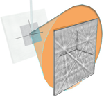 |
EBSD FacilityElectron backscattered diffraction (EBSD) maps are obtained with an Oxford Instruments Nordlys detector. SDI participates in the Jackson School EBSD facility. An EBSD is currently installed on our Zeiss SEM. Lab contact: Esti Ukar Key references: Ukar, E., Laubach, S.E., Marrett, R., 2016, Quartz c-axis orientation patterns in fracture cement as a measure of fracture opening rate and a validation tool for fracture pattern models: Geosphere, v. 12, no. 2, p. 400–438, doi: 10.1130/GES01213.1. | view at publisher Fall, A., Ukar, E., Laubach, S.E., 2016, Origin and timing of Dauphiné twins in quartz cement in fractured sandstones from diagenetic environments: insight from fluid inclusions. Tectonophysics doi.org/10.1016/j.tecto.2016.08.014 | view at publisher |
We also maintain a suite of transmitted light microscopes. For other JSG facilities and equipment available to the project, see the JSG Facilities page.
These facilities available to the Initiative include environmental SEMs, micro CT, atomic force microscopy (AFM), and a wide range of capabilities for geochemical and isotopic analysis.
Additional testing is routinely conducted in our labs at Petroleum & Geosystems Engineering. For more information about current rock mechanics and analog research and test capabilities contact Jon Olson.
For our field methods and a description of our Schmidt hammers, drills, and drones for outcrop imaging, see the Publications page.
Information on project software and field and laboratory methods
Information about the SEM CL Facility


