
CT Scans Revolutionize Fossil Preparation
November 22, 2010
The same year that Francis Whitney taught the university’s first paleontology course, 1909, the American Museum of Natural History in New York published a landmark paper titled “Modern laboratory methods in vertebrate paleontology.” Among other things, the article described best practices for fossil preparation, the art of removing fossils from the rock encasing them. When Matt Brown first came to the university in April 2009 to run the fossil preparation lab, he would open a drawer and find tools still in use that were highlighted in the article.
“Paleontology hasn’t changed much in 100 years,” he says. “Those tools are still very effective, but there are a lot more techniques developed in other industries over the last 20 to 50 years that we’ve been slow to apply to the prep lab.”
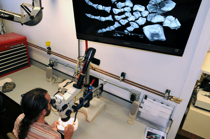
Brown is working hard to change that. He has started sending fossil specimens to the university’s CT lab for scanning before he prepares them. The scans, essentially three dimensional x-rays, serve as a guide to preparing the fossils more safely and efficiently. Back at his workbench, with a hand on a specimen and the other on a tool, Brown glances up at a CT image on a computer screen to see what lies beneath the surface.
“Most of the time, you have to go painfully slowly and treat it as if there could be bone beneath each chisel stroke,” he says. “With CT, I can move quickly through an area with no bone and then slow down as I approach bone. It essentially gives you X-ray vision.”
By some estimates, it can take 10 to 100 hours of preparation for each hour collecting a specimen in the field. Brown notes you have to hold the specimen in just a certain way to be in the field of focus of your microscope and it’s hard to hold your hands, shoulders and head in a comfortable way.
“You spend a lot of time hunched over clutching a tool that’s vibrating and trying to cradle a fragile bone in your other hand,” he says. “So the less time you have to spend on a specimen, the less stress on your body.”
Sometimes, the preparators are opening up a huge block collected decades earlier and jacketed in plaster. Documentation on what might be inside is often thin to nonexistent. With nothing else to guide them, CT scans can provide valuable clues.
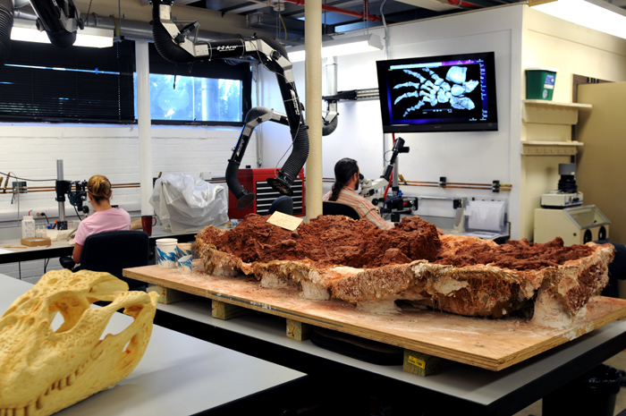
Perhaps all a researcher needs is a tooth from a specimen to identify the species. With CT scans, preparators can go right to the spot, remove a tooth and leave the rest of the specimen unprepared. It saves time that could be used preparing other specimens. And because the best way to preserve fossils is to leave them encased in rock, this targeted approach is better from a conservation standpoint.
Fossils can be damaged during the preparation process or even years later. Imagine a student pulls open an old dusty drawer and the bottom falls out, smashing the carefully prepared fossil on the floor.
“If we have the CT data set, at least we have a digital representation of what it looked like before that happened,” he says.
by Marc Airhart
Also See:
High-Resolution X-ray CT Facility
For more information about the Jackson School contact J.B. Bird at jbird@jsg.utexas.edu, 512-232-9623.