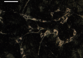
Figure 1. Dark field image of mesenchymal stem cells (MSCs) labeled with gold nanoparticles. Scale bar = 100 µm.
Laura Ricles, Seung Yun Nam, Stanislav Emelianov, and Laura Suggs (Dept. of Biomedical Engineering)
Cardiovascular diseases are the leading cause of death globally. Current research is focused on using stem cell-based therapy for cardiovascular diseases in order to promote neovascularization and rescue ischemic tissue. However, a major challenge to the development of these cellular therapies is the lack of efficient stem cell tracking methods in order to monitor the extent of cell engraftment, mechanisms of vascular regeneration, and participation and role of administered stem cells in vascular repair. To address these issues, we monitor and track mesenchymal stem cells (MSCs) in vivo following implantation. Specifically, we monitor blood vessel growth kinetics and functionin vivo using combined ultrasound/photoacoustic imaging in order to track MSCs labeled with gold nanoparticle contrast agents. In order to perform long-term tracking of MSCs, it is essential that gold nanoparticles are still localized within the cells over time and have not been sufficiently diluted due to cell division. ICP-MS provides a quantitative assessment of nanoparticle concentration within MSCs over time, thus providing us with information related to the feasibility of long-term cell tracking.

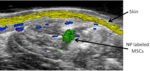
Figure 2. Spectroscopic photoacoustic imaging of nanoparticle labeled MSCs (green) injected into the lateral gastrocnemius. Skin (yellow) and oxygenated (red) and deoxygenated (blue) hemoglobin are also shown.
Figure publications:
- Ricles LM, Nam SY, Sokolov K., Emelianov SY, Sugs LI. 2011. Int J Nanomedicine 6: 407-16.
- Nam SY, Ricles LM, Suggs LJ, Emelianov SY. 2012. PLoS One 7: e37267
© 2024 Jackson School of Geosciences, The University of Texas at Austin

